


 Foundational characteristics of cancer include proliferation, angiogenesis, migration, evasion of apoptosis, and cellular immortality. Find key markers for these cellular processes and antibodies to detect them.
 The SUMOplot™ Analysis Program predicts and scores sumoylation sites in your protein. SUMOylation is a post-translational modification involved in various cellular processes, such as nuclear-cytosolic transport, transcriptional regulation, apoptosis, protein stability, response to stress, and progression through the cell cycle.
 The Autophagy Receptor Motif Plotter predicts and scores autophagy receptor binding sites in your protein. Identifying proteins connected to this pathway is critical to understanding the role of autophagy in physiological as well as pathological processes such as development, differentiation, neurodegenerative diseases, stress, infection, and cancer.
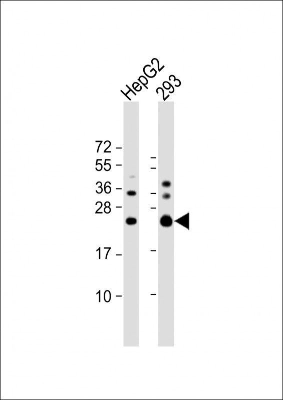
RAB5C Antibody (C-term)
Affinity Purified Rabbit Polyclonal Antibody (Pab)
- SPECIFICATION
- CITATIONS
- PROTOCOLS
- BACKGROUND

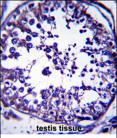
Application
| IHC-P, WB, E |
|---|---|
| Primary Accession | P51148 |
| Other Accession | Q58DS9, NP_004574.2, NP_958842.1 |
| Reactivity | Human |
| Predicted | Bovine |
| Host | Rabbit |
| Clonality | Polyclonal |
| Isotype | Rabbit IgG |
| Calculated MW | 23483 Da |
| Antigen Region | 178-207 aa |
| Gene ID | 5878 |
|---|---|
| Other Names | Ras-related protein Rab-5C, L1880, RAB5L, RAB5C, RABL |
| Target/Specificity | This RAB5C antibody is generated from rabbits immunized with a KLH conjugated synthetic peptide between 178-207 amino acids from the C-terminal region of human RAB5C. |
| Dilution | IHC-P~~1:10~50 WB~~1:1000 E~~Use at an assay dependent concentration. |
| Format | Purified polyclonal antibody supplied in PBS with 0.09% (W/V) sodium azide. This antibody is purified through a protein A column, followed by peptide affinity purification. |
| Storage | Maintain refrigerated at 2-8°C for up to 2 weeks. For long term storage store at -20°C in small aliquots to prevent freeze-thaw cycles. |
| Precautions | RAB5C Antibody (C-term) is for research use only and not for use in diagnostic or therapeutic procedures. |
| Name | RAB5C (HGNC:9785) |
|---|---|
| Synonyms | RABL |
| Function | The small GTPases Rab are key regulators of intracellular membrane trafficking, from the formation of transport vesicles to their fusion with membranes. Rabs cycle between an inactive GDP-bound form and an active GTP-bound form that is able to recruit to membranes different sets of downstream effectors directly responsible for vesicle formation, movement, tethering and fusion. |
| Cellular Location | Cell membrane {ECO:0000250|UniProtKB:P20339}; Lipid-anchor {ECO:0000250|UniProtKB:P20339}; Cytoplasmic side {ECO:0000250|UniProtKB:P20339}. Early endosome membrane {ECO:0000250|UniProtKB:P20339}; Lipid-anchor {ECO:0000250|UniProtKB:P20339}. Melanosome. Note=Identified by mass spectrometry in melanosome fractions from stage I to stage IV |

Thousands of laboratories across the world have published research that depended on the performance of antibodies from Abcepta to advance their research. Check out links to articles that cite our products in major peer-reviewed journals, organized by research category.
info@abcepta.com, and receive a free "I Love Antibodies" mug.

Provided below are standard protocols that you may find useful for product applications.
Background
Members of the Rab protein family are small GTPases of the Ras superfamily that are thought to ensure fidelity in the process of docking and/or fusion of vesicles with their correct acceptor compartment (Han et al., 1996 [PubMed 8646882]).
References
Ewing, R.M., et al. Mol. Syst. Biol. 3, 89 (2007) :
Chi, A., et al. J. Proteome Res. 5(11):3135-3144(2006)
Merithew, E., et al. J. Biol. Chem. 278(10):8494-8500(2003)
Clemens, D.L., et al. Infect. Immun. 68(5):2671-2684(2000)
Bucci, C., et al. Biochem. Biophys. Res. Commun. 258(3):657-662(1999)
If you have used an Abcepta product and would like to share how it has performed, please click on the "Submit Review" button and provide the requested information. Our staff will examine and post your review and contact you if needed.
If you have any additional inquiries please email technical services at tech@abcepta.com.
Ordering Information
Other Products
Shipping Information
