


 Foundational characteristics of cancer include proliferation, angiogenesis, migration, evasion of apoptosis, and cellular immortality. Find key markers for these cellular processes and antibodies to detect them.
 The SUMOplot™ Analysis Program predicts and scores sumoylation sites in your protein. SUMOylation is a post-translational modification involved in various cellular processes, such as nuclear-cytosolic transport, transcriptional regulation, apoptosis, protein stability, response to stress, and progression through the cell cycle.
 The Autophagy Receptor Motif Plotter predicts and scores autophagy receptor binding sites in your protein. Identifying proteins connected to this pathway is critical to understanding the role of autophagy in physiological as well as pathological processes such as development, differentiation, neurodegenerative diseases, stress, infection, and cancer.
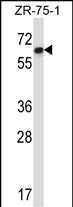
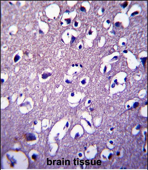
DCTN4 Antibody (N-term)
Affinity Purified Rabbit Polyclonal Antibody (Pab)
- SPECIFICATION
- CITATIONS
- PROTOCOLS
- BACKGROUND

Application
| IHC-P, WB, E |
|---|---|
| Primary Accession | Q9UJW0 |
| Other Accession | Q9QUR2, Q8CBY8, NP_057305.1, A0A4X1TB62 |
| Reactivity | Human |
| Predicted | Mouse, Pig, Rat |
| Host | Rabbit |
| Clonality | Polyclonal |
| Isotype | Rabbit IgG |
| Calculated MW | 52337 Da |
| Antigen Region | 1-30 aa |
| Gene ID | 51164 |
|---|---|
| Other Names | Dynactin subunit 4, Dyn4, Dynactin subunit p62, DCTN4 |
| Target/Specificity | This DCTN4 antibody is generated from rabbits immunized with a KLH conjugated synthetic peptide between 1-30 amino acids from the N-terminal region of human DCTN4. |
| Dilution | IHC-P~~1:10~50 WB~~1:1000 E~~Use at an assay dependent concentration. |
| Format | Purified polyclonal antibody supplied in PBS with 0.09% (W/V) sodium azide. This antibody is purified through a protein A column, followed by peptide affinity purification. |
| Storage | Maintain refrigerated at 2-8°C for up to 2 weeks. For long term storage store at -20°C in small aliquots to prevent freeze-thaw cycles. |
| Precautions | DCTN4 Antibody (N-term) is for research use only and not for use in diagnostic or therapeutic procedures. |
| Name | DCTN4 (HGNC:15518) |
|---|---|
| Function | Part of the dynactin complex that activates the molecular motor dynein for ultra-processive transport along microtubules. |
| Cellular Location | Cytoplasm, cytoskeleton. Cytoplasm, cytoskeleton, microtubule organizing center, centrosome. Cytoplasm, cytoskeleton, stress fiber {ECO:0000250|UniProtKB:Q9QUR2}. Cytoplasm, cell cortex {ECO:0000250|UniProtKB:Q9QUR2}. Cytoplasm, myofibril, sarcomere {ECO:0000250|UniProtKB:Q8CBY8}. Note=Has a punctate cytoplasmic distribution as well as centrosomal distribution typical of dynactin (PubMed:10671518). Overexpression in cultured mammalian cells revealed colocalization with cortical actin, stress fibers, and focal adhesion sites, sites of potential interaction between microtubules and the cell cortex (By similarity). In skeletal muscles, costamere localization requires the presence of ANK2 (By similarity) {ECO:0000250|UniProtKB:Q8CBY8, ECO:0000250|UniProtKB:Q9QUR2, ECO:0000269|PubMed:10671518} |

Thousands of laboratories across the world have published research that depended on the performance of antibodies from Abcepta to advance their research. Check out links to articles that cite our products in major peer-reviewed journals, organized by research category.
info@abcepta.com, and receive a free "I Love Antibodies" mug.

Provided below are standard protocols that you may find useful for product applications.
Background
DCTN4 could have a dual role in dynein targeting and in ACTR1A/Arp1 subunit of dynactin pointed-end capping. Could be involved in ACTR1A pointed-end binding and in additional roles in linking dynein and dynactin to the cortical cytoskeleton.
References
Ayalon, G., et al. Cell 135(7):1189-1200(2008)
Lim, C.M., et al. J. Biol. Chem. 281(20):14006-14014(2006)
Boultwood, J., et al. Genomics 66(1):26-34(2000)
Karki, S., et al. J. Biol. Chem. 275(7):4834-4839(2000)
Bingham, J.B., et al. Curr. Biol. 9(4):223-226(1999)
If you have used an Abcepta product and would like to share how it has performed, please click on the "Submit Review" button and provide the requested information. Our staff will examine and post your review and contact you if needed.
If you have any additional inquiries please email technical services at tech@abcepta.com.
Ordering Information
Shipping Information
