


 Foundational characteristics of cancer include proliferation, angiogenesis, migration, evasion of apoptosis, and cellular immortality. Find key markers for these cellular processes and antibodies to detect them.
 The SUMOplot™ Analysis Program predicts and scores sumoylation sites in your protein. SUMOylation is a post-translational modification involved in various cellular processes, such as nuclear-cytosolic transport, transcriptional regulation, apoptosis, protein stability, response to stress, and progression through the cell cycle.
 The Autophagy Receptor Motif Plotter predicts and scores autophagy receptor binding sites in your protein. Identifying proteins connected to this pathway is critical to understanding the role of autophagy in physiological as well as pathological processes such as development, differentiation, neurodegenerative diseases, stress, infection, and cancer.
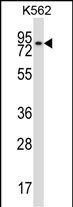
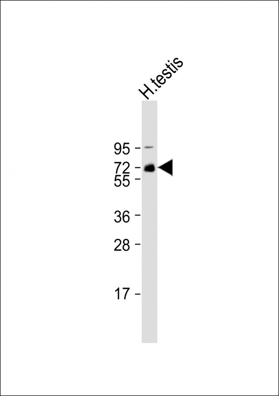
REC8 Antibody (Center)
Affinity Purified Rabbit Polyclonal Antibody (Pab)
- SPECIFICATION
- CITATIONS: 1
- PROTOCOLS
- BACKGROUND

Application
| WB, E |
|---|---|
| Primary Accession | O95072 |
| Other Accession | NP_005123.2, NP_001041670.1 |
| Reactivity | Human |
| Host | Rabbit |
| Clonality | Polyclonal |
| Isotype | Rabbit IgG |
| Calculated MW | 62614 Da |
| Antigen Region | 194-222 aa |
| Gene ID | 9985 |
|---|---|
| Other Names | Meiotic recombination protein REC8 homolog, Cohesin Rec8p, REC8, REC8L1 |
| Target/Specificity | This REC8 antibody is generated from rabbits immunized with a KLH conjugated synthetic peptide between 194-222 amino acids from the Central region of human REC8. |
| Dilution | WB~~1:1000 E~~Use at an assay dependent concentration. |
| Format | Purified polyclonal antibody supplied in PBS with 0.09% (W/V) sodium azide. This antibody is purified through a protein A column, followed by peptide affinity purification. |
| Storage | Maintain refrigerated at 2-8°C for up to 2 weeks. For long term storage store at -20°C in small aliquots to prevent freeze-thaw cycles. |
| Precautions | REC8 Antibody (Center) is for research use only and not for use in diagnostic or therapeutic procedures. |
| Name | REC8 |
|---|---|
| Synonyms | REC8L1 |
| Function | Required during meiosis for separation of sister chromatids and homologous chromosomes. Proteolytic cleavage of REC8 on chromosome arms by separin during anaphase I allows for homologous chromosome separation in meiosis I and cleavage of REC8 on centromeres during anaphase II allows for sister chromatid separation in meiosis II (By similarity). |
| Cellular Location | Nucleus {ECO:0000250|UniProtKB:Q6AYJ4}. Chromosome {ECO:0000250|UniProtKB:Q6AYJ4}. Chromosome, centromere {ECO:0000250|UniProtKB:Q6AYJ4}. Note=In meiotic chromosomes, localized along axial elements in prophase from the leptotene to diplotene stages. At later prophase stages, diakinesis and metaphase I, localized along interstitial axes of chromosomes including both centromere and arm regions. No longer detected in arm regions in anaphase I but persists on centromere regions until metaphase II. Localized to centromeres and spindle poles in endopolyploid tumor cells {ECO:0000250|UniProtKB:Q6AYJ4} |
| Tissue Location | Expressed in testis and thymus. Expressed in the B- cell lines WI-L2-NS and Namalwa (at protein level) |

Provided below are standard protocols that you may find useful for product applications.
Background
This gene encodes a member of the kleisin family of SMC (structural maintenance of chromosome) protein partners. The protein localizes to the axial elements of chromosomes during meiosis in both oocytes and spermatocytes. In the mouse, the homologous protein is a key component of the meiotic cohesion complex, which regulates sister chromatid cohesion and recombination between homologous chromosomes. Multiple alternatively spliced variants, encoding the same protein, have been found for this gene.
References
Aston, K.I., et al. Hum. Reprod. 25(6):1383-1397(2010)
Erenpreisa, J., et al. Exp. Cell Res. 315(15):2593-2603(2009)
Griffin, J., et al. Syst Biol Reprod Med 54(3):163-165(2008)
Xu, H., et al. Dev. Cell 8(6):949-961(2005)
Prieto, I., et al. Chromosome Res. 12(3):197-213(2004)
If you have used an Abcepta product and would like to share how it has performed, please click on the "Submit Review" button and provide the requested information. Our staff will examine and post your review and contact you if needed.
If you have any additional inquiries please email technical services at tech@abcepta.com.
Ordering Information
Other Products
Shipping Information
