


 Foundational characteristics of cancer include proliferation, angiogenesis, migration, evasion of apoptosis, and cellular immortality. Find key markers for these cellular processes and antibodies to detect them.
 The SUMOplot™ Analysis Program predicts and scores sumoylation sites in your protein. SUMOylation is a post-translational modification involved in various cellular processes, such as nuclear-cytosolic transport, transcriptional regulation, apoptosis, protein stability, response to stress, and progression through the cell cycle.
 The Autophagy Receptor Motif Plotter predicts and scores autophagy receptor binding sites in your protein. Identifying proteins connected to this pathway is critical to understanding the role of autophagy in physiological as well as pathological processes such as development, differentiation, neurodegenerative diseases, stress, infection, and cancer.
TIAM2 Antibody (N-term)
Affinity Purified Rabbit Polyclonal Antibody (Pab)
- SPECIFICATION
- CITATIONS
- PROTOCOLS
- BACKGROUND

Application
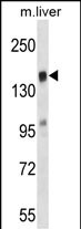
| WB, E |
|---|---|
| Primary Accession | Q8IVF5 |
| Other Accession | Q6ZPF3 |
| Reactivity | Mouse |
| Predicted | Human |
| Host | Rabbit |
| Clonality | Polyclonal |
| Isotype | Rabbit IgG |
| Calculated MW | 190103 Da |
| Antigen Region | 357-385 aa |
| Gene ID | 26230 |
|---|---|
| Other Names | T-lymphoma invasion and metastasis-inducing protein 2, TIAM-2, SIF and TIAM1-like exchange factor, TIAM2, KIAA2016, STEF |
| Target/Specificity | This TIAM2 antibody is generated from rabbits immunized with a KLH conjugated synthetic peptide between 357-385 amino acids of human TIAM2. |
| Dilution | WB~~1:1000 E~~Use at an assay dependent concentration. |
| Format | Purified polyclonal antibody supplied in PBS with 0.09% (W/V) sodium azide. This antibody is purified through a protein A column, followed by peptide affinity purification. |
| Storage | Maintain refrigerated at 2-8°C for up to 2 weeks. For long term storage store at -20°C in small aliquots to prevent freeze-thaw cycles. |
| Precautions | TIAM2 Antibody (N-term) is for research use only and not for use in diagnostic or therapeutic procedures. |
| Name | TIAM2 |
|---|---|
| Synonyms | KIAA2016, STEF |
| Function | Modulates the activity of RHO-like proteins and connects extracellular signals to cytoskeletal activities. Acts as a GDP- dissociation stimulator protein that stimulates the GDP-GTP exchange activity of RHO-like GTPases and activates them. Mediates extracellular laminin signals to activate Rac1, contributing to neurite growth. Involved in lamellipodial formation and advancement of the growth cone of embryonic hippocampal neurons. Promotes migration of neurons in the cerebral cortex. When overexpressed, induces membrane ruffling accompanied by the accumulation of actin filaments along the altered plasma membrane (By similarity). Activates specifically RAC1, but not CDC42 and RHOA. |
| Cellular Location | Cytoplasm {ECO:0000250|UniProtKB:Q6ZPF3}. Cell projection, lamellipodium {ECO:0000250|UniProtKB:Q6ZPF3}. Cell projection, filopodium {ECO:0000250|UniProtKB:Q6ZPF3}. Cell projection, growth cone {ECO:0000250|UniProtKB:Q6ZPF3}. Cell projection, neuron projection {ECO:0000250|UniProtKB:Q6ZPF3}. Perikaryon {ECO:0000250|UniProtKB:Q6ZPF3} |
| Tissue Location | Expressed in the occipital, frontal and temporal lobes, cerebellum, putamen and testis. |

Thousands of laboratories across the world have published research that depended on the performance of antibodies from Abcepta to advance their research. Check out links to articles that cite our products in major peer-reviewed journals, organized by research category.
info@abcepta.com, and receive a free "I Love Antibodies" mug.

Provided below are standard protocols that you may find useful for product applications.
Background
This gene encodes a guanine nucleotide exchange factor. A highly similar mouse protein specifically activates ras-related C3 botulinum substrate 1, converting this Rho-like guanosine triphosphatase (GTPase) from a guanosine diphosphate-bound inactive state to a guanosine triphosphate-bound active state. The encoded protein may play a role in neural cell development. Alternatively spliced transcript variants encoding different isoforms have been described.
References
Rose, J.E., et al. Mol. Med. 16 (7-8), 247-253 (2010) :
Rabizadeh, S., et al. Cytokine Growth Factor Rev. 14 (3-4), 225-239 (2003) :
Salehi, A.H., et al. J. Biol. Chem. 277(50):48043-48050(2002)
Yoshizawa, M., et al. Mech. Dev. 113(1):65-68(2002)
Harrington, A.W., et al. J. Neurosci. 22(1):156-166(2002)
If you have used an Abcepta product and would like to share how it has performed, please click on the "Submit Review" button and provide the requested information. Our staff will examine and post your review and contact you if needed.
If you have any additional inquiries please email technical services at tech@abcepta.com.
Ordering Information
Other Products
Shipping Information
