


 Foundational characteristics of cancer include proliferation, angiogenesis, migration, evasion of apoptosis, and cellular immortality. Find key markers for these cellular processes and antibodies to detect them.
 The SUMOplot™ Analysis Program predicts and scores sumoylation sites in your protein. SUMOylation is a post-translational modification involved in various cellular processes, such as nuclear-cytosolic transport, transcriptional regulation, apoptosis, protein stability, response to stress, and progression through the cell cycle.
 The Autophagy Receptor Motif Plotter predicts and scores autophagy receptor binding sites in your protein. Identifying proteins connected to this pathway is critical to understanding the role of autophagy in physiological as well as pathological processes such as development, differentiation, neurodegenerative diseases, stress, infection, and cancer.
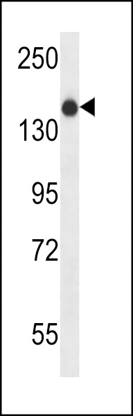
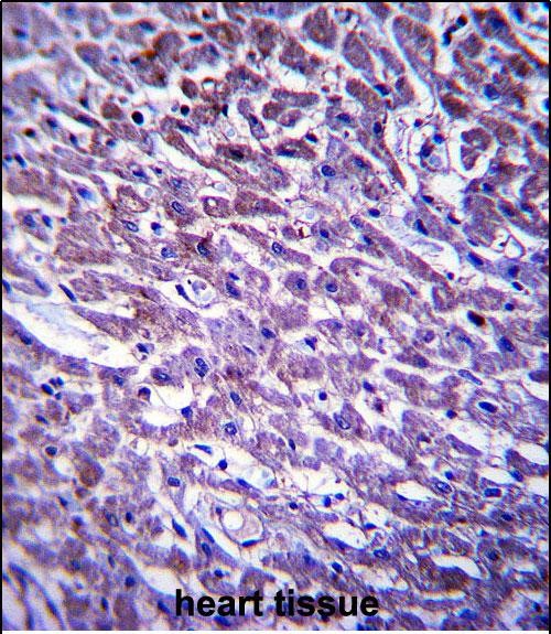
XIRP1 Antibody (C-term)
Affinity Purified Rabbit Polyclonal Antibody (Pab)
- SPECIFICATION
- CITATIONS
- PROTOCOLS
- BACKGROUND

Application
| WB, IHC-P, E |
|---|---|
| Primary Accession | Q702N8 |
| Other Accession | NP_919269.2 |
| Reactivity | Human |
| Host | Rabbit |
| Clonality | Polyclonal |
| Isotype | Rabbit IgG |
| Calculated MW | 198561 Da |
| Antigen Region | 1338-1367 aa |
| Gene ID | 165904 |
|---|---|
| Other Names | Xin actin-binding repeat-containing protein 1, Cardiomyopathy-associated protein 1, XIRP1, CMYA1, XIN |
| Target/Specificity | This XIRP1 antibody is generated from rabbits immunized with a KLH conjugated synthetic peptide between 1338-1367 amino acids from the C-terminal region of human XIRP1. |
| Dilution | WB~~1:1000 IHC-P~~1:10~50 E~~Use at an assay dependent concentration. |
| Format | Purified polyclonal antibody supplied in PBS with 0.09% (W/V) sodium azide. This antibody is purified through a protein A column, followed by peptide affinity purification. |
| Storage | Maintain refrigerated at 2-8°C for up to 2 weeks. For long term storage store at -20°C in small aliquots to prevent freeze-thaw cycles. |
| Precautions | XIRP1 Antibody (C-term) is for research use only and not for use in diagnostic or therapeutic procedures. |
| Name | XIRP1 (HGNC:14301) |
|---|---|
| Function | Protects actin filaments from depolymerization (PubMed:15454575). Required for correct cardiac intercalated disk ultrastructure via maintenance of cell-cell adhesion stability, and as a result maintains cardiac organ morphology, conductance and heart beat rhythm (By similarity). Required for development of normal skeletal muscle morphology and muscle fiber type composition (By similarity). Plays a role in regulating muscle satellite cell activation and survival, as a result promotes muscle fiber recovery from injury and fatigue (By similarity). |
| Cellular Location | Cell junction, adherens junction. Cell junction, desmosome {ECO:0000250|UniProtKB:Q5PZ43}. Note=Colocalizes with actin stress fibers. |
| Tissue Location | Expressed in skeletal muscle at areas of Z-disk disruption in a longitudinal pattern spanning one or more sarcomeres (at protein level). [Isoform B]: Expressed in the heart (at protein level). |

Thousands of laboratories across the world have published research that depended on the performance of antibodies from Abcepta to advance their research. Check out links to articles that cite our products in major peer-reviewed journals, organized by research category.
info@abcepta.com, and receive a free "I Love Antibodies" mug.

Provided below are standard protocols that you may find useful for product applications.
Background
XIRP1 protects actin filaments from depolymerization.
References
Claeys, K.G., et al. Acta Neuropathol. 117(3):293-307(2009)
van der Ven, P.F., et al. Exp. Cell Res. 312(11):2154-2167(2006)
Pacholsky, D., et al. J. Cell. Sci. 117 (PT 22), 5257-5268 (2004) :
Sinn, H.W., et al. Dev. Dyn. 225(1):1-13(2002)
Wang, D.Z., et al. Development 126(6):1281-1294(1999)
If you have used an Abcepta product and would like to share how it has performed, please click on the "Submit Review" button and provide the requested information. Our staff will examine and post your review and contact you if needed.
If you have any additional inquiries please email technical services at tech@abcepta.com.
Ordering Information
Other Products
Shipping Information
