


 Foundational characteristics of cancer include proliferation, angiogenesis, migration, evasion of apoptosis, and cellular immortality. Find key markers for these cellular processes and antibodies to detect them.
 The SUMOplot™ Analysis Program predicts and scores sumoylation sites in your protein. SUMOylation is a post-translational modification involved in various cellular processes, such as nuclear-cytosolic transport, transcriptional regulation, apoptosis, protein stability, response to stress, and progression through the cell cycle.
 The Autophagy Receptor Motif Plotter predicts and scores autophagy receptor binding sites in your protein. Identifying proteins connected to this pathway is critical to understanding the role of autophagy in physiological as well as pathological processes such as development, differentiation, neurodegenerative diseases, stress, infection, and cancer.
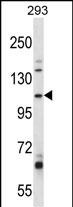
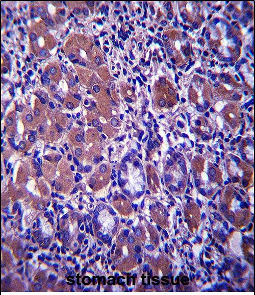
B4GALNT3 Antibody (Center)
Affinity Purified Rabbit Polyclonal Antibody (Pab)
- SPECIFICATION
- CITATIONS
- PROTOCOLS
- BACKGROUND

Application
| IHC-P, WB, E |
|---|---|
| Primary Accession | Q6L9W6 |
| Other Accession | NP_775864.3 |
| Reactivity | Human |
| Host | Rabbit |
| Clonality | Polyclonal |
| Isotype | Rabbit IgG |
| Calculated MW | 114975 Da |
| Antigen Region | 555-584 aa |
| Gene ID | 283358 |
|---|---|
| Other Names | Beta-1, 4-N-acetylgalactosaminyltransferase 3, B4GalNAcT3, Beta4GalNAc-T3, Beta4GalNAcT3, Beta-1, 4-N-acetylgalactosaminyltransferase III, N-acetyl-beta-glucosaminyl-glycoprotein 4-beta-N-acetylgalactosaminyltransferase 2, NGalNAc-T2, B4GALNT3 |
| Target/Specificity | This B4GALNT3 antibody is generated from rabbits immunized with a KLH conjugated synthetic peptide between 555-584 amino acids from the Central region of human B4GALNT3. |
| Dilution | IHC-P~~1:10~50 WB~~1:1000 E~~Use at an assay dependent concentration. |
| Format | Purified polyclonal antibody supplied in PBS with 0.09% (W/V) sodium azide. This antibody is purified through a protein A column, followed by peptide affinity purification. |
| Storage | Maintain refrigerated at 2-8°C for up to 2 weeks. For long term storage store at -20°C in small aliquots to prevent freeze-thaw cycles. |
| Precautions | B4GALNT3 Antibody (Center) is for research use only and not for use in diagnostic or therapeutic procedures. |
| Name | B4GALNT3 |
|---|---|
| Function | Transfers N-acetylgalactosamine (GalNAc) from UDP-GalNAc to N-acetylglucosamine-beta-benzyl with a beta-1,4-linkage to form N,N'- diacetyllactosediamine, GalNAc-beta-1,4-GlcNAc structures in N-linked glycans and probably O-linked glycans. Mediates the N,N'- diacetyllactosediamine formation on gastric mucosa. |
| Cellular Location | Golgi apparatus, Golgi stack membrane; Single-pass type II membrane protein. Note=Localizes to apical Golgi |
| Tissue Location | Highly expressed in testis, colon and stomach. Weakly expressed in other tissues. |

Thousands of laboratories across the world have published research that depended on the performance of antibodies from Abcepta to advance their research. Check out links to articles that cite our products in major peer-reviewed journals, organized by research category.
info@abcepta.com, and receive a free "I Love Antibodies" mug.

Provided below are standard protocols that you may find useful for product applications.
Background
B4GALNT3 transfers N-acetylgalactosamine (GalNAc) onto glucosyl residues to form N,N-prime-diacetyllactosediamine (LacdiNAc, or LDN), a unique terminal structure of cell surface N-glycans (Ikehara et al., 2006 [PubMed 16728562]).[supplied by OMIM].
References
Rose, J. Phd, et al. Mol. Med. (2010) In press :
Arking, D.E., et al. PLoS ONE 5 (3), E9879 (2010) :
Miller, E., et al. J. Biol. Chem. 283(4):1985-1991(2008)
Huang, J., et al. Mol. Cancer Res. 5(6):543-552(2007)
Ikehara, Y., et al. Glycobiology 16(9):777-785(2006)
If you have used an Abcepta product and would like to share how it has performed, please click on the "Submit Review" button and provide the requested information. Our staff will examine and post your review and contact you if needed.
If you have any additional inquiries please email technical services at tech@abcepta.com.
Ordering Information
Other Products
Shipping Information
