


 Foundational characteristics of cancer include proliferation, angiogenesis, migration, evasion of apoptosis, and cellular immortality. Find key markers for these cellular processes and antibodies to detect them.
 The SUMOplot™ Analysis Program predicts and scores sumoylation sites in your protein. SUMOylation is a post-translational modification involved in various cellular processes, such as nuclear-cytosolic transport, transcriptional regulation, apoptosis, protein stability, response to stress, and progression through the cell cycle.
 The Autophagy Receptor Motif Plotter predicts and scores autophagy receptor binding sites in your protein. Identifying proteins connected to this pathway is critical to understanding the role of autophagy in physiological as well as pathological processes such as development, differentiation, neurodegenerative diseases, stress, infection, and cancer.
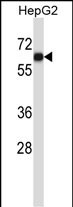
AMIGO1 Antibody (C-term)
Affinity Purified Rabbit Polyclonal Antibody (Pab)
- SPECIFICATION
- CITATIONS
- PROTOCOLS
- BACKGROUND

Application
| WB, E |
|---|---|
| Primary Accession | Q86WK6 |
| Other Accession | Q80ZD7, Q80ZD8, NP_065754.2 |
| Reactivity | Human |
| Predicted | Mouse, Rat |
| Host | Rabbit |
| Clonality | Polyclonal |
| Isotype | Rabbit IgG |
| Calculated MW | 55239 Da |
| Antigen Region | 386-415 aa |
| Gene ID | 57463 |
|---|---|
| Other Names | Amphoterin-induced protein 1, AMIGO-1, Alivin-2, AMIGO1 (HGNC:20824) |
| Target/Specificity | This AMIGO1 antibody is generated from rabbits immunized with a KLH conjugated synthetic peptide between 386-415 amino acids from the C-terminal region of human AMIGO1. |
| Dilution | WB~~1:1000 E~~Use at an assay dependent concentration. |
| Format | Purified polyclonal antibody supplied in PBS with 0.09% (W/V) sodium azide. This antibody is purified through a protein A column, followed by peptide affinity purification. |
| Storage | Maintain refrigerated at 2-8°C for up to 2 weeks. For long term storage store at -20°C in small aliquots to prevent freeze-thaw cycles. |
| Precautions | AMIGO1 Antibody (C-term) is for research use only and not for use in diagnostic or therapeutic procedures. |
| Name | AMIGO1 (HGNC:20824) |
|---|---|
| Function | Promotes growth and fasciculation of neurites from cultured hippocampal neurons. May be involved in fasciculation as well as myelination of developing neural axons. May have a role in regeneration as well as neural plasticity in the adult nervous system. May mediate homophilic as well as heterophilic cell-cell interaction and contribute to signal transduction through its intracellular domain. Assembled with KCNB1 modulates the gating characteristics of the delayed rectifier voltage-dependent potassium channel KCNB1. |
| Cellular Location | Cell membrane {ECO:0000250|UniProtKB:Q80ZD8}; Single-pass type I membrane protein {ECO:0000250|UniProtKB:Q80ZD8} Perikaryon {ECO:0000250|UniProtKB:Q80ZD8}. Cell projection, dendrite {ECO:0000250|UniProtKB:Q80ZD8}. Cell projection, axon {ECO:0000250|UniProtKB:Q80ZD7}. Note=Colocalizes with KCNB1 at high- density somatodendritic clusters on the surface of hippocampal and cortical neurons. Associated with axons of neuronal cells {ECO:0000250|UniProtKB:Q80ZD7, ECO:0000250|UniProtKB:Q80ZD8} |

Thousands of laboratories across the world have published research that depended on the performance of antibodies from Abcepta to advance their research. Check out links to articles that cite our products in major peer-reviewed journals, organized by research category.
info@abcepta.com, and receive a free "I Love Antibodies" mug.

Provided below are standard protocols that you may find useful for product applications.
Background
AMIGO1 promotes growth and fasciculation of neurites from cultured hippocampal neurons. May be involved in fasciculation as well as myelination of developing neural axons. May have a role in regeneration as well as neural plasticity in the adult nervous system. May mediate homophilic as well as heterophilic cell-cell interaction and contribute to signal transduction through its intracellular domain (By similarity).
References
Kottgen, A., et al. Nat. Genet. 42(5):376-384(2010)
Lamesch, P., et al. Genomics 89(3):307-315(2007)
Kuja-Panula, J., et al. J. Cell Biol. 160(6):963-973(2003)
If you have used an Abcepta product and would like to share how it has performed, please click on the "Submit Review" button and provide the requested information. Our staff will examine and post your review and contact you if needed.
If you have any additional inquiries please email technical services at tech@abcepta.com.
Ordering Information
Other Products
Shipping Information
