


 Foundational characteristics of cancer include proliferation, angiogenesis, migration, evasion of apoptosis, and cellular immortality. Find key markers for these cellular processes and antibodies to detect them.
 The SUMOplot™ Analysis Program predicts and scores sumoylation sites in your protein. SUMOylation is a post-translational modification involved in various cellular processes, such as nuclear-cytosolic transport, transcriptional regulation, apoptosis, protein stability, response to stress, and progression through the cell cycle.
 The Autophagy Receptor Motif Plotter predicts and scores autophagy receptor binding sites in your protein. Identifying proteins connected to this pathway is critical to understanding the role of autophagy in physiological as well as pathological processes such as development, differentiation, neurodegenerative diseases, stress, infection, and cancer.
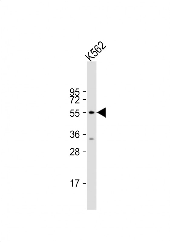
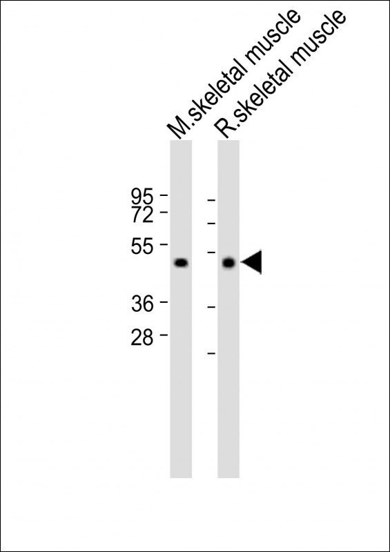
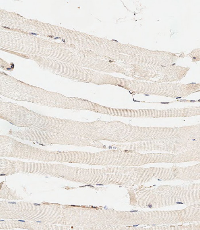
HYAL4 Antibody (Center)
Affinity Purified Rabbit Polyclonal Antibody (Pab)
- SPECIFICATION
- CITATIONS
- PROTOCOLS
- BACKGROUND

Application
| IHC-P-Leica, WB, E |
|---|---|
| Primary Accession | Q2M3T9 |
| Other Accession | NP_036401.2 |
| Reactivity | Human, Mouse, Rat |
| Host | Rabbit |
| Clonality | Polyclonal |
| Isotype | Rabbit IgG |
| Calculated MW | 54249 Da |
| Antigen Region | 143-172 aa |
| Gene ID | 23553 |
|---|---|
| Other Names | Hyaluronidase-4, Hyal-4, Chondroitin sulfate endo-beta-N-acetylgalactosaminidase, Chondroitin sulfate hydrolase, CSHY, Hyaluronoglucosaminidase-4, HYAL4 |
| Target/Specificity | This HYAL4 antibody is generated from rabbits immunized with a KLH conjugated synthetic peptide between 143-172 amino acids from the Central region of human HYAL4. |
| Dilution | IHC-P-Leica~~1:500 WB~~1:2000 E~~Use at an assay dependent concentration. |
| Format | Purified polyclonal antibody supplied in PBS with 0.09% (W/V) sodium azide. This antibody is purified through a protein A column, followed by peptide affinity purification. |
| Storage | Maintain refrigerated at 2-8°C for up to 2 weeks. For long term storage store at -20°C in small aliquots to prevent freeze-thaw cycles. |
| Precautions | HYAL4 Antibody (Center) is for research use only and not for use in diagnostic or therapeutic procedures. |
| Name | HYAL4 |
|---|---|
| Function | Endo-hyaluronidase that degrades hyaluronan to smaller oligosaccharide fragments. Also has chondroitin sulfate hydrolase activity, The best substrate being the galactosaminidic linkage in the sequence of a trisulfated tetrasaccharide. |
| Cellular Location | Membrane; Multi-pass membrane protein |
| Tissue Location | Detected in placenta and skeletal muscle. |

Thousands of laboratories across the world have published research that depended on the performance of antibodies from Abcepta to advance their research. Check out links to articles that cite our products in major peer-reviewed journals, organized by research category.
info@abcepta.com, and receive a free "I Love Antibodies" mug.

Provided below are standard protocols that you may find useful for product applications.
Background
This gene encodes a protein which is similar in structure to hyaluronidases. Hyaluronidases intracellularly degrade hyaluronan, one of the major glycosaminoglycans of the extracellular matrix. Hyaluronan is thought to be involved in cell proliferation, migration and differentiation. However, this protein has not yet been shown to have hyaluronidase activity. [provided by RefSeq].
References
Rose, J.E., et al. Mol. Med. 16 (7-8), 247-253 (2010) :
Kaneiwa, T., et al. Glycobiology 20(3):300-309(2010)
Fiszer-Szafarz, B., et al. J. Biochem. Biophys. Methods 45(2):103-116(2000)
Csoka, A.B., et al. Genomics 60(3):356-361(1999)
If you have used an Abcepta product and would like to share how it has performed, please click on the "Submit Review" button and provide the requested information. Our staff will examine and post your review and contact you if needed.
If you have any additional inquiries please email technical services at tech@abcepta.com.
Ordering Information
Other Products
Shipping Information
