


 Foundational characteristics of cancer include proliferation, angiogenesis, migration, evasion of apoptosis, and cellular immortality. Find key markers for these cellular processes and antibodies to detect them.
 The SUMOplot™ Analysis Program predicts and scores sumoylation sites in your protein. SUMOylation is a post-translational modification involved in various cellular processes, such as nuclear-cytosolic transport, transcriptional regulation, apoptosis, protein stability, response to stress, and progression through the cell cycle.
 The Autophagy Receptor Motif Plotter predicts and scores autophagy receptor binding sites in your protein. Identifying proteins connected to this pathway is critical to understanding the role of autophagy in physiological as well as pathological processes such as development, differentiation, neurodegenerative diseases, stress, infection, and cancer.
RANGAP1 Antibody (N-term)
Affinity Purified Rabbit Polyclonal Antibody (Pab)
- SPECIFICATION
- CITATIONS: 1
- PROTOCOLS
- BACKGROUND

Application
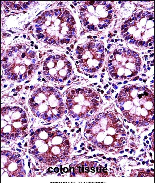
| IHC-P, WB, E |
|---|---|
| Primary Accession | P46060 |
| Other Accession | NP_002874.1 |
| Reactivity | Human |
| Host | Rabbit |
| Clonality | Polyclonal |
| Isotype | Rabbit IgG |
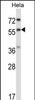
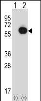
| Calculated MW | 63542 Da |
| Antigen Region | 20-49 aa |
| Gene ID | 5905 |
|---|---|
| Other Names | Ran GTPase-activating protein 1, RanGAP1, RANGAP1, KIAA1835, SD |
| Target/Specificity | This RANGAP1 antibody is generated from rabbits immunized with a KLH conjugated synthetic peptide between 20-49 amino acids from the N-terminal region of human RANGAP1. |
| Dilution | IHC-P~~1:10~50 WB~~1:1000 E~~Use at an assay dependent concentration. |
| Format | Purified polyclonal antibody supplied in PBS with 0.09% (W/V) sodium azide. This antibody is purified through a protein A column, followed by peptide affinity purification. |
| Storage | Maintain refrigerated at 2-8°C for up to 2 weeks. For long term storage store at -20°C in small aliquots to prevent freeze-thaw cycles. |
| Precautions | RANGAP1 Antibody (N-term) is for research use only and not for use in diagnostic or therapeutic procedures. |
| Name | RANGAP1 |
|---|---|
| Synonyms | KIAA1835, SD |
| Function | GTPase activator for RAN (PubMed:16428860, PubMed:8146159, PubMed:8896452). Converts cytoplasmic GTP-bound RAN to GDP-bound RAN, which is essential for RAN-mediated nuclear import and export (PubMed:27160050, PubMed:8896452). Mediates dissociation of cargo from nuclear export complexes containing XPO1, RAN and RANBP2 after nuclear export (PubMed:27160050). |
| Cellular Location | Cytoplasm. Nucleus, nucleoplasm. Nucleus envelope. Chromosome, centromere, kinetochore. Cytoplasm, cytoskeleton, spindle. Note=Cytoplasmic during interphase Detected at the nuclear envelope during interphase (PubMed:11854305, PubMed:15037602). Targeted to the nuclear pores after sumoylation (PubMed:11854305). During mitosis, associates with mitotic spindles, but is essentially not detected at the spindle poles (PubMed:11854305, PubMed:15037602). Association with kinetochores appears soon after nuclear envelope breakdown and persists until late anaphase (PubMed:11854305). Mitotic location also requires sumoylation (PubMed:11854305). |
| Tissue Location | Highly expressed in brain, thymus and testis. |

Provided below are standard protocols that you may find useful for product applications.
Background
RanGAP1, is a homodimeric 65-kD polypeptide that specifically induces the GTPase activity of RAN, but not of RAS by over 1,000-fold. RanGAP1 is the immediate antagonist of RCC1, a regulator molecule that keeps RAN in the active, GTP-bound state. The RANGAP1 gene encodes a 587-amino acid polypeptide. The sequence is unrelated to that of GTPase activators for other RAS-related proteins, but is 88% identical to Fug1, the murine homolog of yeast Rna1p. RanGAP1 and RCC1 control RAN-dependent transport between the nucleus and cytoplasm. RanGAP1 is a key regulator of the RAN GTP/GDP cycle.
References
Zhang, J., et al. Biochem. Biophys. Res. Commun. 375(2):252-255(2008)
Zuccolo, M., et al. EMBO J. 26(7):1853-1864(2007)
Ewing, R.M., et al. Mol. Syst. Biol. 3, 89 (2007) :
Vertegaal, A.C., et al. Mol. Cell Proteomics 5(12):2298-2310(2006)
Vertegaal, A.C., et al. Mol. Cell Proteomics 5(12):2298-2310(2006)
If you have used an Abcepta product and would like to share how it has performed, please click on the "Submit Review" button and provide the requested information. Our staff will examine and post your review and contact you if needed.
If you have any additional inquiries please email technical services at tech@abcepta.com.
Ordering Information
Other Products
Shipping Information
