


 Foundational characteristics of cancer include proliferation, angiogenesis, migration, evasion of apoptosis, and cellular immortality. Find key markers for these cellular processes and antibodies to detect them.
 The SUMOplot™ Analysis Program predicts and scores sumoylation sites in your protein. SUMOylation is a post-translational modification involved in various cellular processes, such as nuclear-cytosolic transport, transcriptional regulation, apoptosis, protein stability, response to stress, and progression through the cell cycle.
 The Autophagy Receptor Motif Plotter predicts and scores autophagy receptor binding sites in your protein. Identifying proteins connected to this pathway is critical to understanding the role of autophagy in physiological as well as pathological processes such as development, differentiation, neurodegenerative diseases, stress, infection, and cancer.
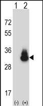
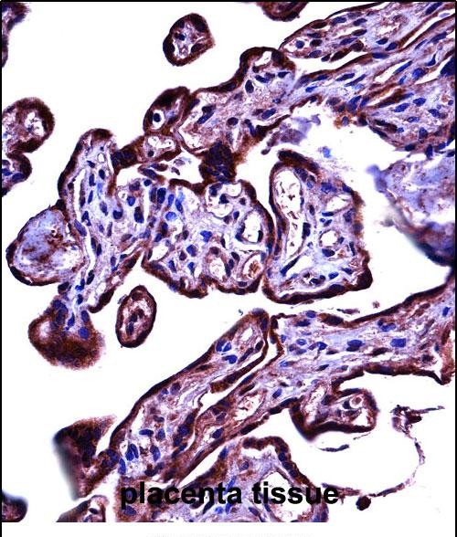
CEACAM3 Antibody (C-term)
Affinity Purified Rabbit Polyclonal Antibody (Pab)
- SPECIFICATION
- CITATIONS
- PROTOCOLS
- BACKGROUND

Application
| IHC-P, WB, E |
|---|---|
| Primary Accession | P40198 |
| Other Accession | NP_001806.2 |
| Reactivity | Human |
| Host | Rabbit |
| Clonality | Polyclonal |
| Isotype | Rabbit IgG |
| Calculated MW | 27091 Da |
| Antigen Region | 163-191 aa |
| Gene ID | 1084 |
|---|---|
| Other Names | Carcinoembryonic antigen-related cell adhesion molecule 3, Carcinoembryonic antigen CGM1, CD66d, CEACAM3, CD66D, CGM1 |
| Target/Specificity | This CEACAM3 antibody is generated from rabbits immunized with a KLH conjugated synthetic peptide between 163-191 amino acids from the C-terminal region of human CEACAM3. |
| Dilution | IHC-P~~1:10~50 WB~~1:1000 E~~Use at an assay dependent concentration. |
| Format | Purified polyclonal antibody supplied in PBS with 0.09% (W/V) sodium azide. This antibody is purified through a protein A column, followed by peptide affinity purification. |
| Storage | Maintain refrigerated at 2-8°C for up to 2 weeks. For long term storage store at -20°C in small aliquots to prevent freeze-thaw cycles. |
| Precautions | CEACAM3 Antibody (C-term) is for research use only and not for use in diagnostic or therapeutic procedures. |
| Name | CEACAM3 (HGNC:1815) |
|---|---|
| Function | Major granulocyte receptor mediating recognition and efficient opsonin-independent phagocytosis of CEACAM-binding microorganisms, including Neissiria, Moxarella and Haemophilus species, thus playing an important role in the clearance of pathogens by the innate immune system. Responsible for RAC1 stimulation in the course of pathogen phagocytosis. |
| Cellular Location | Membrane; Single-pass type I membrane protein. |
| Tissue Location | CGM1a, the predominant CGM1 transcript, is granulocyte-specific. Not detected out of the granulocytic lineage, such as monocytes, lymphocytes, spleen, testis, colon, brain, liver, pancreas, thymus, ovary, placenta, skeletal muscle, prostate, small intestine, heart, lung and kidney. |

Thousands of laboratories across the world have published research that depended on the performance of antibodies from Abcepta to advance their research. Check out links to articles that cite our products in major peer-reviewed journals, organized by research category.
info@abcepta.com, and receive a free "I Love Antibodies" mug.

Provided below are standard protocols that you may find useful for product applications.
Background
This gene encodes a member of the family of carcinoembryonic antigen-related cell adhesion molecules (CEACAMs), which are used by several bacterial pathogens to bind and invade host cells. The encoded transmembrane protein directs phagocytosis of several bacterial species that is dependent on the small GTPase Rac. It is thought to serve an important role in controlling human-specific pathogens by the innate immune system. Alternatively spliced transcript variants have been described, but their biological validity has not been determined.
References
Rose, J.E., et al. Mol. Med. 16 (7-8), 247-253 (2010) :
Tsavaris, N., et al. J Chemother 21(6):673-680(2009)
Skubitz, K.M., et al. J Transl Med 6, 78 (2008) :
Stern-Ginossar, N., et al. J. Immunol. 179(7):4424-4434(2007)
Ali, C.W., et al. J Gastrointest Cancer 38 (2-4), 108-114 (2007) :
If you have used an Abcepta product and would like to share how it has performed, please click on the "Submit Review" button and provide the requested information. Our staff will examine and post your review and contact you if needed.
If you have any additional inquiries please email technical services at tech@abcepta.com.
Ordering Information
Shipping Information
