


 Foundational characteristics of cancer include proliferation, angiogenesis, migration, evasion of apoptosis, and cellular immortality. Find key markers for these cellular processes and antibodies to detect them.
 The SUMOplot™ Analysis Program predicts and scores sumoylation sites in your protein. SUMOylation is a post-translational modification involved in various cellular processes, such as nuclear-cytosolic transport, transcriptional regulation, apoptosis, protein stability, response to stress, and progression through the cell cycle.
 The Autophagy Receptor Motif Plotter predicts and scores autophagy receptor binding sites in your protein. Identifying proteins connected to this pathway is critical to understanding the role of autophagy in physiological as well as pathological processes such as development, differentiation, neurodegenerative diseases, stress, infection, and cancer.
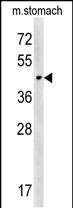
CHST14 Antibody (Center)
Affinity Purified Rabbit Polyclonal Antibody (Pab)
- SPECIFICATION
- CITATIONS
- PROTOCOLS
- BACKGROUND

Application
| WB, E |
|---|---|
| Primary Accession | Q8NCH0 |
| Other Accession | Q80V53, NP_569735.1 |
| Reactivity | Mouse |
| Host | Rabbit |
| Clonality | Polyclonal |
| Isotype | Rabbit IgG |
| Calculated MW | 42997 Da |
| Antigen Region | 178-205 aa |
| Gene ID | 113189 |
|---|---|
| Other Names | Carbohydrate sulfotransferase 14, Dermatan 4-sulfotransferase 1, D4ST-1, hD4ST1, CHST14, D4ST1 |
| Target/Specificity | This CHST14 antibody is generated from rabbits immunized with a KLH conjugated synthetic peptide between 178-205 amino acids from the Central region of human CHST14. |
| Dilution | WB~~1:1000 E~~Use at an assay dependent concentration. |
| Format | Purified polyclonal antibody supplied in PBS with 0.09% (W/V) sodium azide. This antibody is purified through a protein A column, followed by peptide affinity purification. |
| Storage | Maintain refrigerated at 2-8°C for up to 2 weeks. For long term storage store at -20°C in small aliquots to prevent freeze-thaw cycles. |
| Precautions | CHST14 Antibody (Center) is for research use only and not for use in diagnostic or therapeutic procedures. |
| Name | CHST14 |
|---|---|
| Synonyms | D4ST1 |
| Function | Catalyzes the transfer of sulfate to position 4 of the N- acetylgalactosamine (GalNAc) residue of dermatan sulfate. Plays a pivotal role in the formation of 4-0-sulfated IdoA blocks in dermatan sulfate. Transfers sulfate to the C-4 hydroxyl of beta1,4-linked GalNAc that is substituted with an alpha-linked iduronic acid (IdoUA) at the C-3 hydroxyl. Transfers sulfate more efficiently to GalNAc residues in -IdoUA-GalNAc-IdoUA- than in -GlcUA-GalNAc-GlcUA-sequences. Has preference for partially desulfated dermatan sulfate. Addition of sulfate to GalNAc may occur immediately after epimerization of GlcUA to IdoUA. Appears to have an important role in the formation of the cerebellar neural network during postnatal brain development. |
| Cellular Location | Golgi apparatus membrane; Single- pass type II membrane protein |
| Tissue Location | Widely expressed. Expressed at high level in pituitary gland, placenta, uterus and thyroid |

Thousands of laboratories across the world have published research that depended on the performance of antibodies from Abcepta to advance their research. Check out links to articles that cite our products in major peer-reviewed journals, organized by research category.
info@abcepta.com, and receive a free "I Love Antibodies" mug.

Provided below are standard protocols that you may find useful for product applications.
Background
This gene encodes a member of the HNK-1 family of sulfotransferases. The encoded protein transfers sulfate to the C-4 hydroxyl of N-acetylgalactosamine residues in dermatan sulfate. Mutations in this gene have been associated with adducted thumb-clubfoot syndrome.
References
Miyake, N., et al. Hum. Mutat. 31(8):966-974(2010)
Dundar, M., et al. Am. J. Hum. Genet. 85(6):873-882(2009)
Pacheco, B., et al. Glycobiology 19(11):1197-1203(2009)
Lamesch, P., et al. Genomics 89(3):307-315(2007)
Mikami, T., et al. J. Biol. Chem. 278(38):36115-36127(2003)
If you have used an Abcepta product and would like to share how it has performed, please click on the "Submit Review" button and provide the requested information. Our staff will examine and post your review and contact you if needed.
If you have any additional inquiries please email technical services at tech@abcepta.com.
Ordering Information
Other Products
Shipping Information
