


 Foundational characteristics of cancer include proliferation, angiogenesis, migration, evasion of apoptosis, and cellular immortality. Find key markers for these cellular processes and antibodies to detect them.
 The SUMOplot™ Analysis Program predicts and scores sumoylation sites in your protein. SUMOylation is a post-translational modification involved in various cellular processes, such as nuclear-cytosolic transport, transcriptional regulation, apoptosis, protein stability, response to stress, and progression through the cell cycle.
 The Autophagy Receptor Motif Plotter predicts and scores autophagy receptor binding sites in your protein. Identifying proteins connected to this pathway is critical to understanding the role of autophagy in physiological as well as pathological processes such as development, differentiation, neurodegenerative diseases, stress, infection, and cancer.
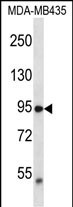
TRPV2 Antibody (N-term)
Affinity Purified Rabbit Polyclonal Antibody (Pab)
- SPECIFICATION
- CITATIONS
- PROTOCOLS
- BACKGROUND

Application
| WB, E |
|---|---|
| Primary Accession | Q9Y5S1 |
| Other Accession | NP_057197.2 |
| Reactivity | Human |
| Host | Rabbit |
| Clonality | Polyclonal |
| Isotype | Rabbit IgG |
| Calculated MW | 85981 Da |
| Antigen Region | 52-81 aa |
| Gene ID | 51393 |
|---|---|
| Other Names | Transient receptor potential cation channel subfamily V member 2, TrpV2, Osm-9-like TRP channel 2, OTRPC2, Vanilloid receptor-like protein 1, VRL-1, TRPV2, VRL |
| Target/Specificity | This TRPV2 antibody is generated from rabbits immunized with a KLH conjugated synthetic peptide between 52-81 amino acids from the N-terminal region of human TRPV2. |
| Dilution | WB~~1:1000 E~~Use at an assay dependent concentration. |
| Format | Purified polyclonal antibody supplied in PBS with 0.09% (W/V) sodium azide. This antibody is purified through a protein A column, followed by peptide affinity purification. |
| Storage | Maintain refrigerated at 2-8°C for up to 2 weeks. For long term storage store at -20°C in small aliquots to prevent freeze-thaw cycles. |
| Precautions | TRPV2 Antibody (N-term) is for research use only and not for use in diagnostic or therapeutic procedures. |
| Name | TRPV2 |
|---|---|
| Synonyms | VRL |
| Function | Calcium-permeable, non-selective cation channel with an outward rectification. Seems to be regulated, at least in part, by IGF1, PDGF and neuropeptide head activator. May transduce physical stimuli in mast cells. Activated by temperatures higher than 52 degrees Celsius; is not activated by vanilloids and acidic pH. |
| Cellular Location | Cell membrane {ECO:0000250|UniProtKB:Q9WTR1}; Multi-pass membrane protein. Cytoplasm {ECO:0000250|UniProtKB:Q9WTR1}. Melanosome. Note=Translocates from the cytoplasm to the plasma membrane upon ligand stimulation (By similarity). Identified by mass spectrometry in melanosome fractions from stage I to stage IV {ECO:0000250|UniProtKB:Q9WTR1} |

Thousands of laboratories across the world have published research that depended on the performance of antibodies from Abcepta to advance their research. Check out links to articles that cite our products in major peer-reviewed journals, organized by research category.
info@abcepta.com, and receive a free "I Love Antibodies" mug.

Provided below are standard protocols that you may find useful for product applications.
Background
This gene encodes an ion channel that is activated by high temperatures above 52 degrees Celsius. The protein may be involved in transduction of high-temperature heat responses in sensory ganglia. It is thought that in other tissues the channel may be activated by stimuli other than heat.
References
Mercado, J., et al. J. Neurosci. 30(40):13338-13347(2010)
Nabissi, M., et al. Carcinogenesis 31(5):794-803(2010)
Monet, M., et al. Cancer Res. 70(3):1225-1235(2010)
Liu, G., et al. Cancer Genet. Cytogenet. 197(1):54-59(2010)
Saunders, C.I., et al. Biochim. Biophys. Acta 1792(10):1019-1026(2009)
If you have used an Abcepta product and would like to share how it has performed, please click on the "Submit Review" button and provide the requested information. Our staff will examine and post your review and contact you if needed.
If you have any additional inquiries please email technical services at tech@abcepta.com.
Ordering Information
Other Products
Shipping Information
