


 Foundational characteristics of cancer include proliferation, angiogenesis, migration, evasion of apoptosis, and cellular immortality. Find key markers for these cellular processes and antibodies to detect them.
 The SUMOplot™ Analysis Program predicts and scores sumoylation sites in your protein. SUMOylation is a post-translational modification involved in various cellular processes, such as nuclear-cytosolic transport, transcriptional regulation, apoptosis, protein stability, response to stress, and progression through the cell cycle.
 The Autophagy Receptor Motif Plotter predicts and scores autophagy receptor binding sites in your protein. Identifying proteins connected to this pathway is critical to understanding the role of autophagy in physiological as well as pathological processes such as development, differentiation, neurodegenerative diseases, stress, infection, and cancer.
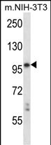
Mouse Ltk Antibody (C-term)
Affinity Purified Rabbit Polyclonal Antibody (Pab)
- SPECIFICATION
- CITATIONS
- PROTOCOLS
- BACKGROUND

Application
| WB, E |
|---|---|
| Primary Accession | P08923 |
| Other Accession | NP_976220.2, NP_996825.2, NP_996824.1 |
| Reactivity | Mouse |
| Host | Rabbit |
| Clonality | Polyclonal |
| Isotype | Rabbit IgG |
| Calculated MW | 94471 Da |
| Antigen Region | 795-822 aa |
| Gene ID | 17005 |
|---|---|
| Other Names | Leukocyte tyrosine kinase receptor, Ltk |
| Target/Specificity | This Mouse Ltk antibody is generated from rabbits immunized with a KLH conjugated synthetic peptide between 795-822 amino acids from the C-terminal region of mouse Ltk. |
| Dilution | WB~~1:1000 E~~Use at an assay dependent concentration. |
| Format | Purified polyclonal antibody supplied in PBS with 0.09% (W/V) sodium azide. This antibody is purified through a protein A column, followed by peptide affinity purification. |
| Storage | Maintain refrigerated at 2-8°C for up to 2 weeks. For long term storage store at -20°C in small aliquots to prevent freeze-thaw cycles. |
| Precautions | Mouse Ltk Antibody (C-term) is for research use only and not for use in diagnostic or therapeutic procedures. |
| Name | Ltk {ECO:0000303|PubMed:2357970} |
|---|---|
| Function | Receptor with a tyrosine-protein kinase activity. Following activation by ALKAL1 or ALKAL2 ligands at the cell surface, transduces an extracellular signal into an intracellular response. Ligand-binding to the extracellular domain induces tyrosine kinase activation, leading to activation of the mitogen-activated protein kinase (MAPK) pathway (By similarity). Phosphorylates almost exclusively at the first tyrosine of the Y-x-x-x-Y-Y motif (By similarity). The exact function of this protein is not known; studies with chimeric proteins demonstrate its ability to promote growth and specifically neurite outgrowth, and cell survival. Involved in regulation of the secretory pathway involving endoplasmic reticulum (ER) export sites (ERESs) and ER to Golgi transport (By similarity). |
| Cellular Location | Cell membrane; Single-pass type I membrane protein [Isoform B]: Endoplasmic reticulum. Note=Retained in the endoplasmic reticulum. |
| Tissue Location | Subsets of lymphoid and neuronal cells. |

Thousands of laboratories across the world have published research that depended on the performance of antibodies from Abcepta to advance their research. Check out links to articles that cite our products in major peer-reviewed journals, organized by research category.
info@abcepta.com, and receive a free "I Love Antibodies" mug.

Provided below are standard protocols that you may find useful for product applications.
Background
The protein encoded by this gene is a member of the ros/insulin receptor family of tyrosine kinases. Tyrosine-specific phosphorylation of proteins is a key to the control of diverse pathways leading to cell growth and differentiation. Four alternatively spliced transcript variants encoding different isoforms have been described for this gene. These transcripts are expressed in a tissue-specific manner in lymphocytes, brain and neuroblastoma cells, and the encoded isoforms exhibit different subcellular localization. The lymphocyte and brain specific variants initiate translation at non-AUG (CUG) start codons.
References
Li, J., et al. J. Biol. Chem. 283(49):34260-34272(2008)
Yu, X., et al. J. Immunol. 177(10):7042-7049(2006)
Li, N., et al. Hum. Mol. Genet. 13(2):171-179(2004)
Thut, C.J., et al. Dev. Biol. 231(1):63-76(2001)
Robinson, D.R., et al. Oncogene 19(49):5548-5557(2000)
If you have used an Abcepta product and would like to share how it has performed, please click on the "Submit Review" button and provide the requested information. Our staff will examine and post your review and contact you if needed.
If you have any additional inquiries please email technical services at tech@abcepta.com.
Ordering Information
Other Products
Shipping Information
