


 Foundational characteristics of cancer include proliferation, angiogenesis, migration, evasion of apoptosis, and cellular immortality. Find key markers for these cellular processes and antibodies to detect them.
 The SUMOplot™ Analysis Program predicts and scores sumoylation sites in your protein. SUMOylation is a post-translational modification involved in various cellular processes, such as nuclear-cytosolic transport, transcriptional regulation, apoptosis, protein stability, response to stress, and progression through the cell cycle.
 The Autophagy Receptor Motif Plotter predicts and scores autophagy receptor binding sites in your protein. Identifying proteins connected to this pathway is critical to understanding the role of autophagy in physiological as well as pathological processes such as development, differentiation, neurodegenerative diseases, stress, infection, and cancer.
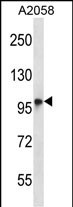
TMC6 Antibody (N-term)
Affinity Purified Rabbit Polyclonal Antibody (Pab)
- SPECIFICATION
- CITATIONS
- PROTOCOLS
- BACKGROUND

Application
| WB, E |
|---|---|
| Primary Accession | Q7Z403 |
| Other Accession | NP_009198.4, NP_001120670.1 |
| Reactivity | Human |
| Host | Rabbit |
| Clonality | Polyclonal |
| Isotype | Rabbit IgG |
| Calculated MW | 90045 Da |
| Antigen Region | 160-188 aa |
| Gene ID | 11322 |
|---|---|
| Other Names | Transmembrane channel-like protein 6, Epidermodysplasia verruciformis protein 1, Protein LAK-4, TMC6, EVER1, EVIN1 |
| Target/Specificity | This TMC6 antibody is generated from rabbits immunized with a KLH conjugated synthetic peptide between 160-188 amino acids from the N-terminal region of human TMC6. |
| Dilution | WB~~1:1000 E~~Use at an assay dependent concentration. |
| Format | Purified polyclonal antibody supplied in PBS with 0.09% (W/V) sodium azide. This antibody is purified through a protein A column, followed by peptide affinity purification. |
| Storage | Maintain refrigerated at 2-8°C for up to 2 weeks. For long term storage store at -20°C in small aliquots to prevent freeze-thaw cycles. |
| Precautions | TMC6 Antibody (N-term) is for research use only and not for use in diagnostic or therapeutic procedures. |
| Name | TMC6 (HGNC:18021) |
|---|---|
| Function | Acts as a regulatory protein involved in the regulation of numerous cellular processes (PubMed:18158319, PubMed:30068544, PubMed:32917726). Together with its homolog TMC8/EVER2, forms a complex with CIB1 in lymphocytes and keratynocytes where TMC6 and TMC8 stabilize CIB1 and reciprocally (PubMed:30068544, PubMed:32917726). Together with TMC8, also forms a complex with and activates zinc transporter ZNT1 at the ER membrane of keratynocytes, thereby facilitating zinc uptake into the ER (PubMed:18158319). Down-regulates the activity of transcription factors induced by zinc and cytokines (PubMed:18158319). Also plays a role in thermal sensation by inhibiting the M-channel (KCNQ2-KCNQ3 channel) current in primary sensory neurons (By similarity). |
| Cellular Location | Endoplasmic reticulum membrane; Multi-pass membrane protein. Golgi apparatus membrane; Multi-pass membrane protein. Nucleus membrane; Multi-pass membrane protein. Note=Localizes to the ER, Golgi and nucleus membranes in keratinocytes. |
| Tissue Location | Expressed in placenta, prostate, testis, activated T-lymphocytes and lymphokine-activated killer (LAK) lymphocytes {ECO:0000269|PubMed:12906855, ECO:0000269|Ref.3} |

Thousands of laboratories across the world have published research that depended on the performance of antibodies from Abcepta to advance their research. Check out links to articles that cite our products in major peer-reviewed journals, organized by research category.
info@abcepta.com, and receive a free "I Love Antibodies" mug.

Provided below are standard protocols that you may find useful for product applications.
Background
Epidermodysplasia verruciformis (EV) is an autosomal recessive dermatosis characterized by abnormal susceptibility to human papillomaviruses (HPVs) and a high rate of progression to squamous cell carcinoma on sun-exposed skin. EV is caused by mutations in either of two adjacent genes located on chromosome 17q25.3. Both of these genes encode integral membrane proteins that localize to the endoplasmic reticulum and are predicted to form transmembrane channels. This gene encodes a transmembrane channel-like protein with 10 transmembrane domains and 2 leucine zipper motifs.
References
McDermott, D.F., et al. Pediatr Dermatol 26(3):306-310(2009)
Lazarczyk, M., et al. J. Exp. Med. 205(1):35-42(2008)
Zuo, Y.G., et al. J. Dermatol. Sci. 44(3):153-159(2006)
Olsen, J.V., et al. Cell 127(3):635-648(2006)
Donfack, J., et al. Int. J. Pediatr. Otorhinolaryngol. 70(7):1235-1240(2006)
If you have used an Abcepta product and would like to share how it has performed, please click on the "Submit Review" button and provide the requested information. Our staff will examine and post your review and contact you if needed.
If you have any additional inquiries please email technical services at tech@abcepta.com.
Ordering Information
Shipping Information
