


 Foundational characteristics of cancer include proliferation, angiogenesis, migration, evasion of apoptosis, and cellular immortality. Find key markers for these cellular processes and antibodies to detect them.
 The SUMOplot™ Analysis Program predicts and scores sumoylation sites in your protein. SUMOylation is a post-translational modification involved in various cellular processes, such as nuclear-cytosolic transport, transcriptional regulation, apoptosis, protein stability, response to stress, and progression through the cell cycle.
 The Autophagy Receptor Motif Plotter predicts and scores autophagy receptor binding sites in your protein. Identifying proteins connected to this pathway is critical to understanding the role of autophagy in physiological as well as pathological processes such as development, differentiation, neurodegenerative diseases, stress, infection, and cancer.
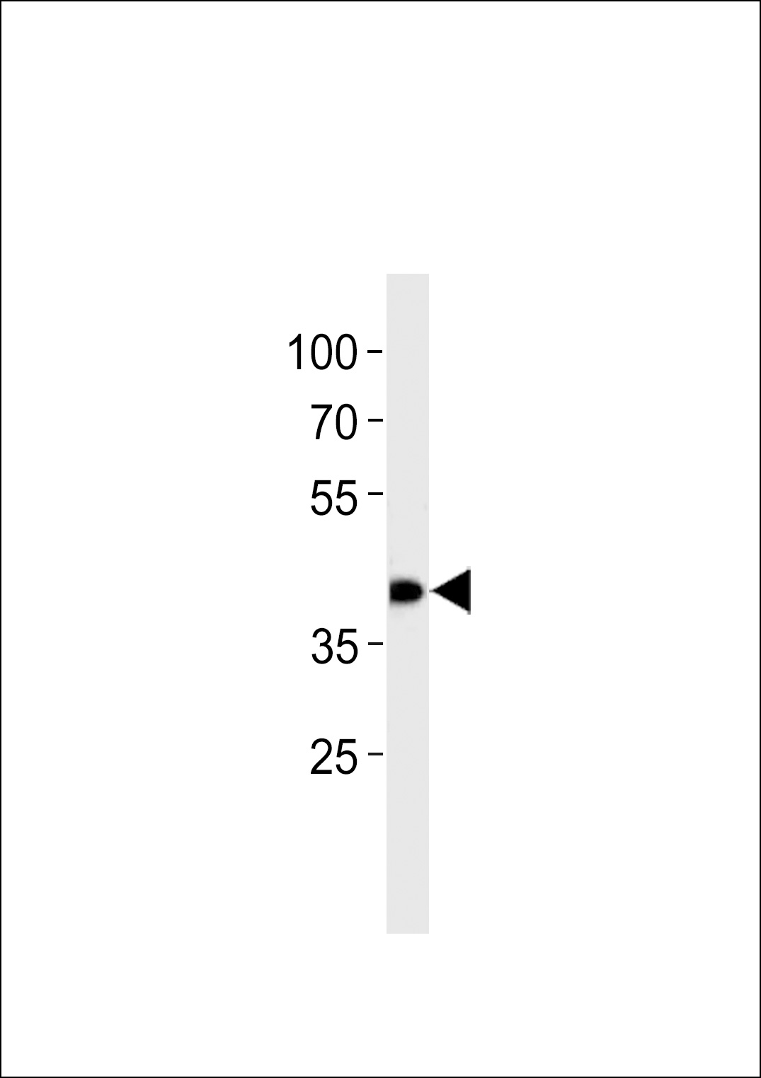
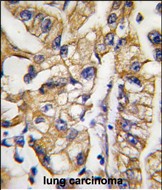
HDBP1 Antibody (Center)
Purified Rabbit Polyclonal Antibody (Pab)
- SPECIFICATION
- CITATIONS: 1
- PROTOCOLS
- BACKGROUND

Application
| WB, IHC-P, E |
|---|---|
| Primary Accession | Q9NR83 |
| Reactivity | Human |
| Host | Rabbit |
| Clonality | Polyclonal |
| Isotype | Rabbit IgG |
| Calculated MW | 41267 Da |
| Antigen Region | 213-242 aa |
| Gene ID | 56731 |
|---|---|
| Other Names | SLC2A4 regulator, GLUT4 enhancer factor, GEF, Huntington disease gene regulatory region-binding protein 1, HDBP-1, SLC2A4RG, HDBP1 |
| Target/Specificity | This HDBP1 antibody is generated from rabbits immunized with a KLH conjugated synthetic peptide between 213-242 amino acids from the Central region of human HDBP1. |
| Dilution | WB~~1:1000 IHC-P~~1:10~50 E~~Use at an assay dependent concentration. |
| Format | Purified polyclonal antibody supplied in PBS with 0.09% (W/V) sodium azide. This antibody is purified through a protein G column, followed by dialysis against PBS. |
| Storage | Maintain refrigerated at 2-8°C for up to 2 weeks. For long term storage store at -20°C in small aliquots to prevent freeze-thaw cycles. |
| Precautions | HDBP1 Antibody (Center) is for research use only and not for use in diagnostic or therapeutic procedures. |
| Name | SLC2A4RG |
|---|---|
| Synonyms | HDBP1 |
| Function | Transcription factor involved in SLC2A4 and HD gene transactivation. Binds to the consensus sequence 5'-GCCGGCG-3'. |
| Cellular Location | Cytoplasm. Nucleus. Note=Shuttles between the cytoplasm and the nucleus |
| Tissue Location | According to PubMed:14630949, expressed in heart, skeletal muscle, liver, kidney and pancreas; undetectable in lung, placenta or brain. According to PubMed:14625278, ubiquitously expressed, with lowest expression in brain and ileum |

Provided below are standard protocols that you may find useful for product applications.
Background
THDBP1 is a nuclear transcription factor involved in the activation of the solute carrier family 2 member 4 gene. This protein interacts with another transcription factor, myocyte enhancer factor 2, to activate transcription of this gene.
References
Jones,M.R., Fertil. Steril. (2008)
McGee,S.L., FASEB J. 20 (2), 348-349 (2006)
Tanaka,K., J. Biol. Chem. 279 (8), 7275-7286 (2004)
If you have used an Abcepta product and would like to share how it has performed, please click on the "Submit Review" button and provide the requested information. Our staff will examine and post your review and contact you if needed.
If you have any additional inquiries please email technical services at tech@abcepta.com.
Ordering Information
Shipping Information
