


 Foundational characteristics of cancer include proliferation, angiogenesis, migration, evasion of apoptosis, and cellular immortality. Find key markers for these cellular processes and antibodies to detect them.
 The SUMOplot™ Analysis Program predicts and scores sumoylation sites in your protein. SUMOylation is a post-translational modification involved in various cellular processes, such as nuclear-cytosolic transport, transcriptional regulation, apoptosis, protein stability, response to stress, and progression through the cell cycle.
 The Autophagy Receptor Motif Plotter predicts and scores autophagy receptor binding sites in your protein. Identifying proteins connected to this pathway is critical to understanding the role of autophagy in physiological as well as pathological processes such as development, differentiation, neurodegenerative diseases, stress, infection, and cancer.
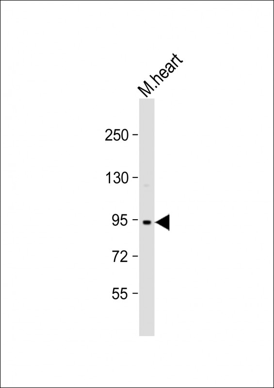
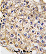
Mouse TRPV3 Antibody (N-term)
Purified Rabbit Polyclonal Antibody (Pab)
- SPECIFICATION
- CITATIONS
- PROTOCOLS
- BACKGROUND

Application
| IHC-P, WB, E |
|---|---|
| Primary Accession | Q8K424 |
| Reactivity | Mouse |
| Host | Rabbit |
| Clonality | Polyclonal |
| Isotype | Rabbit IgG |
| Calculated MW | 90663 Da |
| Antigen Region | 90-119 aa |
| Gene ID | 246788 |
|---|---|
| Other Names | Transient receptor potential cation channel subfamily V member 3, TrpV3, Trpv3 |
| Target/Specificity | This Mouse TRPV3 antibody is generated from rabbits immunized with a KLH conjugated synthetic peptide between 90-119 amino acids from the N-terminal region of mouse TRPV3. |
| Dilution | IHC-P~~1:10~50 WB~~1:1000 E~~Use at an assay dependent concentration. |
| Format | Purified polyclonal antibody supplied in PBS with 0.09% (W/V) sodium azide. This antibody is purified through a protein G column, followed by dialysis against PBS. |
| Storage | Maintain refrigerated at 2-8°C for up to 2 weeks. For long term storage store at -20°C in small aliquots to prevent freeze-thaw cycles. |
| Precautions | Mouse TRPV3 Antibody (N-term) is for research use only and not for use in diagnostic or therapeutic procedures. |
| Name | Trpv3 |
|---|---|
| Function | Non-selective calcium permeant cation channel (PubMed:12016205). It is activated by innocuous (warm) temperatures and shows an increased response at noxious temperatures greater than 39 degrees Celsius (By similarity). Activation exhibits an outward rectification (By similarity). The channel pore can dilate to provide permeability to larger cations (By similarity). May associate with TRPV1 and may modulate its activity (By similarity). Is a negative regulator of hair growth and cycling: TRPV3-coupled signaling suppresses keratinocyte proliferation in hair follicles and induces apoptosis and premature hair follicle regression (catagen) (By similarity). |
| Cellular Location | Cell membrane {ECO:0000250|UniProtKB:Q8NET8}; Multi-pass membrane protein {ECO:0000250|UniProtKB:Q8NET8}. Cytoplasm {ECO:0000250|UniProtKB:Q8NET8}. Lysosome {ECO:0000250|UniProtKB:Q8NET8}. Note=Targeted to lysosome for degradation in a SNX11-dependent manner {ECO:0000250|UniProtKB:Q8NET8} |
| Tissue Location | Expressed in keratinocytes and hair follicles. |

Thousands of laboratories across the world have published research that depended on the performance of antibodies from Abcepta to advance their research. Check out links to articles that cite our products in major peer-reviewed journals, organized by research category.
info@abcepta.com, and receive a free "I Love Antibodies" mug.

Provided below are standard protocols that you may find useful for product applications.
Background
TRPV3 belongs to a family of nonselective cation channels that function in a variety of processes, including temperature sensation and vasoregulation. The thermosensitive members of this family are expressed in subsets of sensory neurons that terminate in the skin, and are activated at distinct physiological temperatures. This channel is activated at temperatures between 22 and 40 degrees C. This gene lies in close proximity to another family member (TRPV1) gene on chromosome 17, and the two encoded proteins are thought to associate with each other to form heteromeric channels.
References
Frederick,J., Biochem. Biophys. Res. Commun. 358 (4), 1058-1064 (2007)
Asakawa,M., J. Invest. Dermatol. 126 (12), 2664-2672 (2006)
If you have used an Abcepta product and would like to share how it has performed, please click on the "Submit Review" button and provide the requested information. Our staff will examine and post your review and contact you if needed.
If you have any additional inquiries please email technical services at tech@abcepta.com.
Ordering Information
Other Products
Shipping Information
