


 Foundational characteristics of cancer include proliferation, angiogenesis, migration, evasion of apoptosis, and cellular immortality. Find key markers for these cellular processes and antibodies to detect them.
 The SUMOplot™ Analysis Program predicts and scores sumoylation sites in your protein. SUMOylation is a post-translational modification involved in various cellular processes, such as nuclear-cytosolic transport, transcriptional regulation, apoptosis, protein stability, response to stress, and progression through the cell cycle.
 The Autophagy Receptor Motif Plotter predicts and scores autophagy receptor binding sites in your protein. Identifying proteins connected to this pathway is critical to understanding the role of autophagy in physiological as well as pathological processes such as development, differentiation, neurodegenerative diseases, stress, infection, and cancer.
TEAD3 Antibody (N-term)
Affinity Purified Rabbit Polyclonal Antibody (Pab)
- SPECIFICATION
- CITATIONS
- PROTOCOLS
- BACKGROUND

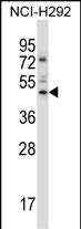
Application
| WB, IHC-P, E |
|---|---|
| Primary Accession | Q99594 |
| Other Accession | P70210, NP_003205.2 |
| Reactivity | Human |
| Predicted | Mouse |
| Host | Rabbit |
| Clonality | Polyclonal |
| Isotype | Rabbit IgG |
| Calculated MW | 48676 Da |
| Antigen Region | 82-110 aa |
| Gene ID | 7005 |
|---|---|
| Other Names | Transcriptional enhancer factor TEF-5, DTEF-1, TEA domain family member 3, TEAD-3, TEAD3, TEAD5, TEF5 |
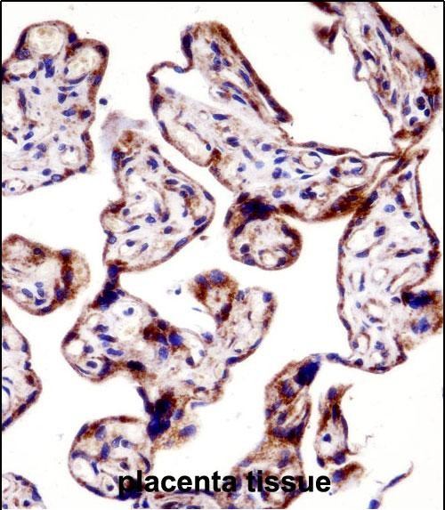
| Target/Specificity | This TEAD3 antibody is generated from rabbits immunized with a KLH conjugated synthetic peptide between 82-110 amino acids from the N-terminal region of human TEAD3. |
| Dilution | WB~~1:1000 IHC-P~~1:10~50 E~~Use at an assay dependent concentration. |
| Format | Purified polyclonal antibody supplied in PBS with 0.09% (W/V) sodium azide. This antibody is purified through a protein A column, followed by peptide affinity purification. |
| Storage | Maintain refrigerated at 2-8°C for up to 2 weeks. For long term storage store at -20°C in small aliquots to prevent freeze-thaw cycles. |
| Precautions | TEAD3 Antibody (N-term) is for research use only and not for use in diagnostic or therapeutic procedures. |
| Name | TEAD3 |
|---|---|
| Synonyms | TEAD5, TEF5 |
| Function | Transcription factor which plays a key role in the Hippo signaling pathway, a pathway involved in organ size control and tumor suppression by restricting proliferation and promoting apoptosis. The core of this pathway is composed of a kinase cascade wherein MST1/MST2, in complex with its regulatory protein SAV1, phosphorylates and activates LATS1/2 in complex with its regulatory protein MOB1, which in turn phosphorylates and inactivates YAP1 oncoprotein and WWTR1/TAZ. Acts by mediating gene expression of YAP1 and WWTR1/TAZ, thereby regulating cell proliferation, migration and epithelial mesenchymal transition (EMT) induction. Binds to multiple functional elements of the human chorionic somatomammotropin-B gene enhancer. |
| Cellular Location | Nucleus. |
| Tissue Location | Preferentially expressed in the placenta. |

Thousands of laboratories across the world have published research that depended on the performance of antibodies from Abcepta to advance their research. Check out links to articles that cite our products in major peer-reviewed journals, organized by research category.
info@abcepta.com, and receive a free "I Love Antibodies" mug.

Provided below are standard protocols that you may find useful for product applications.
Background
This gene product is a member of the transcriptional enhancer factor (TEF) family of transcription factors, which contain the TEA/ATTS DNA-binding domain. It is predominantly expressed in the placenta and is involved in the transactivation of the chorionic somatomammotropin-B gene enhancer. Translation of this protein is initiated at a non-AUG (AUA) start codon. [provided by RefSeq].
References
Zhang, H., et al. J. Biol. Chem. 284(20):13355-13362(2009)
Zhao, B., et al. Genes Dev. 22(14):1962-1971(2008)
Peng, L., et al. Mol. Endocrinol. 18(8):2049-2060(2004)
Mungall, A.J., et al. Nature 425(6960):805-811(2003)
Maeda, T., et al. J. Biol. Chem. 277(27):24346-24352(2002)
If you have used an Abcepta product and would like to share how it has performed, please click on the "Submit Review" button and provide the requested information. Our staff will examine and post your review and contact you if needed.
If you have any additional inquiries please email technical services at tech@abcepta.com.
Ordering Information
Other Products
Shipping Information
