


 Foundational characteristics of cancer include proliferation, angiogenesis, migration, evasion of apoptosis, and cellular immortality. Find key markers for these cellular processes and antibodies to detect them.
 The SUMOplot™ Analysis Program predicts and scores sumoylation sites in your protein. SUMOylation is a post-translational modification involved in various cellular processes, such as nuclear-cytosolic transport, transcriptional regulation, apoptosis, protein stability, response to stress, and progression through the cell cycle.
 The Autophagy Receptor Motif Plotter predicts and scores autophagy receptor binding sites in your protein. Identifying proteins connected to this pathway is critical to understanding the role of autophagy in physiological as well as pathological processes such as development, differentiation, neurodegenerative diseases, stress, infection, and cancer.
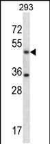
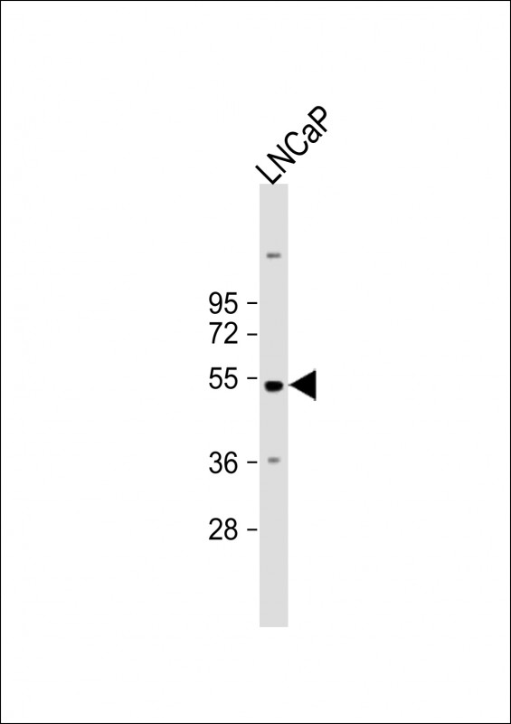
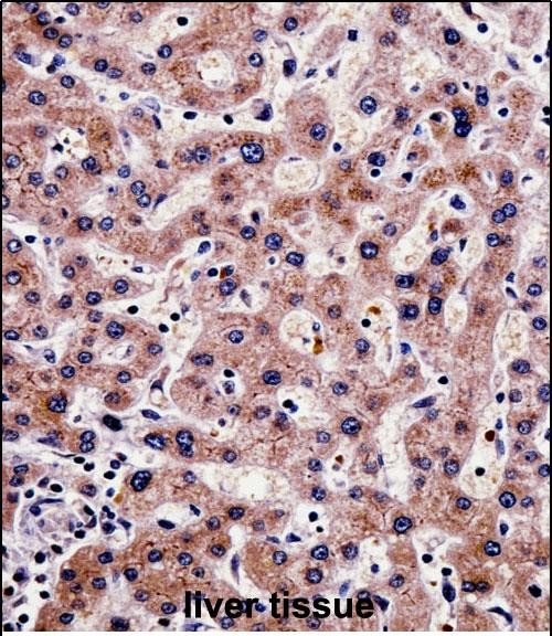
SHMT1 Antibody (N-term)
Affinity Purified Rabbit Polyclonal Antibody (Pab)
- SPECIFICATION
- CITATIONS: 1
- PROTOCOLS
- BACKGROUND

Application
| IHC-P, WB, E |
|---|---|
| Primary Accession | P34896 |
| Other Accession | Q5E9P9, NP_004160.3, NP_683718.1, P35623 |
| Reactivity | Human |
| Predicted | Bovine, Sheep |
| Host | Rabbit |
| Clonality | Polyclonal |
| Isotype | Rabbit IgG |
| Calculated MW | 53083 Da |
| Antigen Region | 19-47 aa |
| Gene ID | 6470 |
|---|---|
| Other Names | Serine hydroxymethyltransferase, cytosolic, SHMT, Glycine hydroxymethyltransferase, Serine methylase, SHMT1 |
| Target/Specificity | This SHMT1 antibody is generated from rabbits immunized with a KLH conjugated synthetic peptide between 19-47 amino acids from the N-terminal region of human SHMT1. |
| Dilution | IHC-P~~1:10~50 WB~~1:1000 E~~Use at an assay dependent concentration. |
| Format | Purified polyclonal antibody supplied in PBS with 0.09% (W/V) sodium azide. This antibody is purified through a protein A column, followed by peptide affinity purification. |
| Storage | Maintain refrigerated at 2-8°C for up to 2 weeks. For long term storage store at -20°C in small aliquots to prevent freeze-thaw cycles. |
| Precautions | SHMT1 Antibody (N-term) is for research use only and not for use in diagnostic or therapeutic procedures. |
| Name | SHMT1 |
|---|---|
| Function | Interconversion of serine and glycine (PubMed:24698160, PubMed:8505317). |
| Cellular Location | Cytoplasm. |

Provided below are standard protocols that you may find useful for product applications.
Background
This gene encodes the cellular form of serine hydroxymethyltransferase, a pyridoxal phosphate-containing enzyme that catalyzes the reversible conversion of serine and tetrahydrofolate to glycine and 5,10-methylene tetrahydrofolate. This reaction provides one carbon units for synthesis of methionine, thymidylate, and purines in the cytoplasm. This gene is located within the Smith-Magenis syndrome region on chromosome 17. Alternative splicing of this gene results in 2 transcript variants encoding 2 different isoforms. Additional transcript variants have been described, but their biological validity has not been determined.
References
Porter, K.E., et al. Environ. Res. 110(6):580-587(2010)
Summers, C.M., et al. Birth Defects Res. Part A Clin. Mol. Teratol. 88(8):679-688(2010)
Vijayakrishnan, J., et al. Haematologica 95(8):1405-1414(2010)
Levine, A.J., et al. Cancer Epidemiol. Biomarkers Prev. 19(7):1812-1821(2010)
Jugessur, A., et al. PLoS ONE 5 (7), E11493 (2010) :
If you have used an Abcepta product and would like to share how it has performed, please click on the "Submit Review" button and provide the requested information. Our staff will examine and post your review and contact you if needed.
If you have any additional inquiries please email technical services at tech@abcepta.com.
Ordering Information
Other Products
Shipping Information
