


 Foundational characteristics of cancer include proliferation, angiogenesis, migration, evasion of apoptosis, and cellular immortality. Find key markers for these cellular processes and antibodies to detect them.
 The SUMOplot™ Analysis Program predicts and scores sumoylation sites in your protein. SUMOylation is a post-translational modification involved in various cellular processes, such as nuclear-cytosolic transport, transcriptional regulation, apoptosis, protein stability, response to stress, and progression through the cell cycle.
 The Autophagy Receptor Motif Plotter predicts and scores autophagy receptor binding sites in your protein. Identifying proteins connected to this pathway is critical to understanding the role of autophagy in physiological as well as pathological processes such as development, differentiation, neurodegenerative diseases, stress, infection, and cancer.
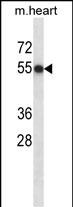
Mouse Csnk1d Antibody (Center)
Affinity Purified Rabbit Polyclonal Antibody (Pab)
- SPECIFICATION
- CITATIONS
- PROTOCOLS
- BACKGROUND

Application
| WB, E |
|---|---|
| Primary Accession | Q9DC28 |
| Other Accession | Q9JMK2, P49674, Q5ZLL1, Q5BP74, Q06486, P48730, P35508, Q6P3K7, Q7T2E3, NP_620690.1 |
| Reactivity | Mouse |
| Predicted | Zebrafish, Bovine, Human, Rat, Xenopus, Chicken |
| Host | Rabbit |
| Clonality | Polyclonal |
| Isotype | Rabbit IgG |
| Calculated MW | 47316 Da |
| Antigen Region | 204-231 aa |
| Gene ID | 104318 |
|---|---|
| Other Names | Casein kinase I isoform delta, CKI-delta, CKId, Tau-protein kinase CSNK1D, Csnk1d, Hckid |
| Target/Specificity | This Mouse Csnk1d antibody is generated from rabbits immunized with a KLH conjugated synthetic peptide between 204-231 amino acids from the Central region of mouse Csnk1d. |
| Dilution | WB~~1:1000 E~~Use at an assay dependent concentration. |
| Format | Purified polyclonal antibody supplied in PBS with 0.09% (W/V) sodium azide. This antibody is purified through a protein A column, followed by peptide affinity purification. |
| Storage | Maintain refrigerated at 2-8°C for up to 2 weeks. For long term storage store at -20°C in small aliquots to prevent freeze-thaw cycles. |
| Precautions | Mouse Csnk1d Antibody (Center) is for research use only and not for use in diagnostic or therapeutic procedures. |
| Name | Csnk1d |
|---|---|
| Synonyms | Hckid |
| Function | Essential serine/threonine-protein kinase that regulates diverse cellular growth and survival processes including Wnt signaling, DNA repair and circadian rhythms. It can phosphorylate a large number of proteins. Casein kinases are operationally defined by their preferential utilization of acidic proteins such as caseins as substrates. Phosphorylates connexin-43/GJA1, MAP1A, SNAPIN, MAPT/TAU, TOP2A, DCK, HIF1A, EIF6, p53/TP53, DVL2, DVL3, ESR1, AIB1/NCOA3, DNMT1, PKD2, YAP1, PER1 and PER2. Central component of the circadian clock. In balance with PP1, determines the circadian period length through the regulation of the speed and rhythmicity of PER1 and PER2 phosphorylation. Controls PER1 and PER2 nuclear transport and degradation. YAP1 phosphorylation promotes its SCF(beta-TRCP) E3 ubiquitin ligase-mediated ubiquitination and subsequent degradation. DNMT1 phosphorylation reduces its DNA-binding activity. Phosphorylation of ESR1 and AIB1/NCOA3 stimulates their activity and coactivation. Phosphorylation of DVL2 and DVL3 regulates WNT3A signaling pathway that controls neurite outgrowth. Phosphorylates NEDD9/HEF1 (PubMed:29191835). EIF6 phosphorylation promotes its nuclear export. Triggers down-regulation of dopamine receptors in the forebrain. Activates DCK in vitro by phosphorylation. TOP2A phosphorylation favors DNA cleavable complex formation. May regulate the formation of the mitotic spindle apparatus in extravillous trophoblast. Modulates connexin-43/GJA1 gap junction assembly by phosphorylation. Probably involved in lymphocyte physiology. Regulates fast synaptic transmission mediated by glutamate. |
| Cellular Location | Cytoplasm. Nucleus. Cytoplasm, cytoskeleton, microtubule organizing center, centrosome. Cytoplasm, perinuclear region. Cell membrane. Cytoplasm, cytoskeleton, spindle. Golgi apparatus Note=Localized at mitotic spindle microtubules, and at the centrosomes and interphase in interphase cells. Recruited to the spindle apparatus and the centrosomes in response to DNA-damage. Correct subcellular localization requires kinase activity (By similarity). |
| Tissue Location | Expressed ubiquitously. However, kinase activity is not uniform, with highest kinase activity in splenocytes |

Thousands of laboratories across the world have published research that depended on the performance of antibodies from Abcepta to advance their research. Check out links to articles that cite our products in major peer-reviewed journals, organized by research category.
info@abcepta.com, and receive a free "I Love Antibodies" mug.

Provided below are standard protocols that you may find useful for product applications.
Background
This gene encodes a member of the casein kinase I (CKI) family of serine/threonine protein kinases. A highly similar human protein regulates an array of cellular processes by influencing the Wnt and hedgehog signaling pathways. The encoded protein may also be involved in the regulation of apoptosis, circadian rhythm, microtubule dynamics, chromosome segregation, and p53-mediated effects on growth. Alternatively spliced transcript variants encoding different isoforms have been described. [provided by RefSeq].
References
Sugiyama, Y., et al. Biochem. J. 427(3):489-497(2010)
Etchegaray, J.P., et al. PLoS ONE 5 (4), E10303 (2010) :
Lee, H., et al. Proc. Natl. Acad. Sci. U.S.A. 106(50):21359-21364(2009)
Martinez, G., et al. Invest. Ophthalmol. Vis. Sci. 50(10):4794-4806(2009)
Isojima, Y., et al. Proc. Natl. Acad. Sci. U.S.A. 106(37):15744-15749(2009)
If you have used an Abcepta product and would like to share how it has performed, please click on the "Submit Review" button and provide the requested information. Our staff will examine and post your review and contact you if needed.
If you have any additional inquiries please email technical services at tech@abcepta.com.
Ordering Information
Other Products
Shipping Information
