


 Foundational characteristics of cancer include proliferation, angiogenesis, migration, evasion of apoptosis, and cellular immortality. Find key markers for these cellular processes and antibodies to detect them.
 The SUMOplot™ Analysis Program predicts and scores sumoylation sites in your protein. SUMOylation is a post-translational modification involved in various cellular processes, such as nuclear-cytosolic transport, transcriptional regulation, apoptosis, protein stability, response to stress, and progression through the cell cycle.
 The Autophagy Receptor Motif Plotter predicts and scores autophagy receptor binding sites in your protein. Identifying proteins connected to this pathway is critical to understanding the role of autophagy in physiological as well as pathological processes such as development, differentiation, neurodegenerative diseases, stress, infection, and cancer.
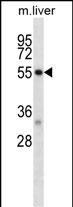
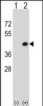
Mouse Dapk2 Antibody (N-term)
Affinity Purified Rabbit Polyclonal Antibody (Pab)
- SPECIFICATION
- CITATIONS
- PROTOCOLS
- BACKGROUND

Application
| WB, E |
|---|---|
| Primary Accession | Q8VDF3 |
| Other Accession | Q9UIK4, NP_034149.2 |
| Reactivity | Human, Mouse |
| Host | Rabbit |
| Clonality | Polyclonal |
| Isotype | Rabbit IgG |
| Calculated MW | 42778 Da |
| Antigen Region | 38-65 aa |
| Gene ID | 13143 |
|---|---|
| Other Names | Death-associated protein kinase 2, DAP kinase 2, DAP-kinase-related protein 1, DRP-1, Dapk2 |
| Target/Specificity | This Mouse Dapk2 antibody is generated from rabbits immunized with a KLH conjugated synthetic peptide between 38-65 amino acids from the N-terminal region of mouse Dapk2. |
| Dilution | WB~~1:1000 E~~Use at an assay dependent concentration. |
| Format | Purified polyclonal antibody supplied in PBS with 0.09% (W/V) sodium azide. This antibody is purified through a protein A column, followed by peptide affinity purification. |
| Storage | Maintain refrigerated at 2-8°C for up to 2 weeks. For long term storage store at -20°C in small aliquots to prevent freeze-thaw cycles. |
| Precautions | Mouse Dapk2 Antibody (N-term) is for research use only and not for use in diagnostic or therapeutic procedures. |
| Name | Dapk2 |
|---|---|
| Function | Calcium/calmodulin-dependent serine/threonine kinase involved in multiple cellular signaling pathways that trigger cell survival, apoptosis, and autophagy. Capable of regulating both type I apoptotic and type II autophagic cell death signals. The former involves caspase activation, chromatin and mitochondrial condensation while the latter involves caspase-independent cell death in conjunction with accumulation of mature autophagic vesicles, plasma membrane blebs, and nuclear condensation without DNA degradation. Mediator of anoikis and a suppressor of beta-catenin-dependent anchorage-independent growth of malignant epithelial cells. May play a role in granulocytic maturation (By similarity). Regulates granulocytes motility by controlling cell spreading and polarization (PubMed:24163421). |
| Cellular Location | Cytoplasm. Cytoplasmic vesicle, autophagosome lumen |
| Tissue Location | Expressed in peritubular interstitial cells of the renal cortex (PubMed:24906443). Isoform 1 is found in the adult brain while isoform 2 is expressed in brains of embryos and young mice (at protein level) (PubMed:21408167). |

Thousands of laboratories across the world have published research that depended on the performance of antibodies from Abcepta to advance their research. Check out links to articles that cite our products in major peer-reviewed journals, organized by research category.
info@abcepta.com, and receive a free "I Love Antibodies" mug.

Provided below are standard protocols that you may find useful for product applications.
Background
Calcium/calmodulin-dependent serine/threonine kinase which acts as a positive regulator of apoptosis (By similarity).
References
Liljander, M., et al. Genet. Res. 91(4):259-265(2009)
Fang, J., et al. Blood 112(3):886-890(2008)
Zambrowicz, B.P., et al. Proc. Natl. Acad. Sci. U.S.A. 100(24):14109-14114(2003)
Shimoda, M., et al. J. Exp. Med. 194(11):1597-1607(2001)
Inbal, B., et al. Mol. Cell. Biol. 20(3):1044-1054(2000)
If you have used an Abcepta product and would like to share how it has performed, please click on the "Submit Review" button and provide the requested information. Our staff will examine and post your review and contact you if needed.
If you have any additional inquiries please email technical services at tech@abcepta.com.
Ordering Information
Other Products
Shipping Information
