


 Foundational characteristics of cancer include proliferation, angiogenesis, migration, evasion of apoptosis, and cellular immortality. Find key markers for these cellular processes and antibodies to detect them.
 The SUMOplot™ Analysis Program predicts and scores sumoylation sites in your protein. SUMOylation is a post-translational modification involved in various cellular processes, such as nuclear-cytosolic transport, transcriptional regulation, apoptosis, protein stability, response to stress, and progression through the cell cycle.
 The Autophagy Receptor Motif Plotter predicts and scores autophagy receptor binding sites in your protein. Identifying proteins connected to this pathway is critical to understanding the role of autophagy in physiological as well as pathological processes such as development, differentiation, neurodegenerative diseases, stress, infection, and cancer.
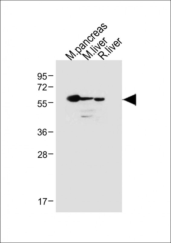
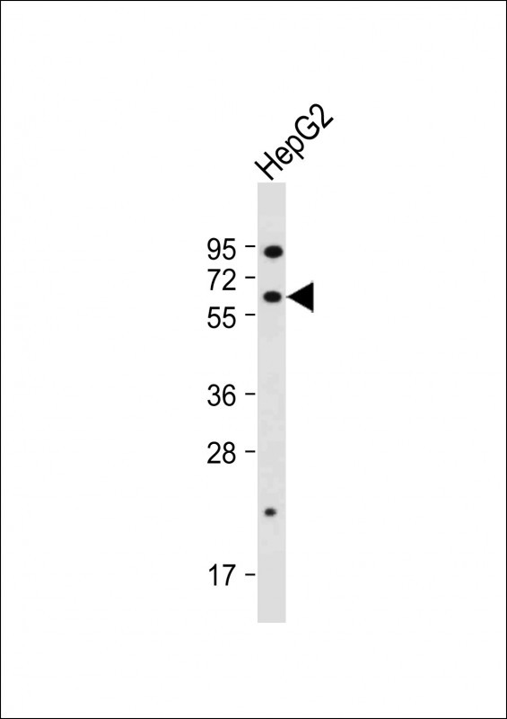
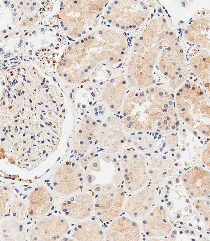
USP30 Antibody (N-term)
Affinity Purified Rabbit Polyclonal Antibody (Pab)
- SPECIFICATION
- CITATIONS
- PROTOCOLS
- BACKGROUND

Application
| IHC-P-Leica, WB, E |
|---|---|
| Primary Accession | Q70CQ3 |
| Other Accession | D3ZPG5, Q3UN04, NP_116052.2 |
| Reactivity | Human, Mouse, Rat |
| Predicted | Mouse, Rat |
| Host | Rabbit |
| Clonality | Polyclonal |
| Isotype | Rabbit IgG |
| Calculated MW | 58503 Da |
| Antigen Region | 156-185 aa |
| Gene ID | 84749 |
|---|---|
| Other Names | Ubiquitin carboxyl-terminal hydrolase 30, Deubiquitinating enzyme 30, Ubiquitin thioesterase 30, Ubiquitin-specific-processing protease 30, Ub-specific protease 30, USP30 |
| Target/Specificity | This USP30 antibody is generated from rabbits immunized with a KLH conjugated synthetic peptide between 156-185 amino acids from the N-terminal region of human USP30. |
| Dilution | IHC-P-Leica~~1:500 WB~~1:2000 E~~Use at an assay dependent concentration. |
| Format | Purified polyclonal antibody supplied in PBS with 0.09% (W/V) sodium azide. This antibody is purified through a protein A column, followed by peptide affinity purification. |
| Storage | Maintain refrigerated at 2-8°C for up to 2 weeks. For long term storage store at -20°C in small aliquots to prevent freeze-thaw cycles. |
| Precautions | USP30 Antibody (N-term) is for research use only and not for use in diagnostic or therapeutic procedures. |
| Name | USP30 (HGNC:20065) |
|---|---|
| Function | Deubiquitinating enzyme tethered to the mitochondrial outer membrane that acts as a key inhibitor of mitophagy by counteracting the action of parkin (PRKN): hydrolyzes ubiquitin attached by parkin on target proteins, such as RHOT1/MIRO1 and TOMM20, thereby blocking parkin's ability to drive mitophagy (PubMed:18287522, PubMed:24896179, PubMed:25527291, PubMed:25621951). Preferentially cleaves 'Lys-6'- and 'Lys-11'-linked polyubiquitin chains, 2 types of linkage that participate in mitophagic signaling (PubMed:25621951). Does not cleave efficiently polyubiquitin phosphorylated at 'Ser-65' (PubMed:25527291). Acts as a negative regulator of mitochondrial fusion by mediating deubiquitination of MFN1 and MFN2 (By similarity). |
| Cellular Location | Mitochondrion outer membrane |
| Tissue Location | Expressed in skeletal muscle, pancreas, liver and kidney. |

Thousands of laboratories across the world have published research that depended on the performance of antibodies from Abcepta to advance their research. Check out links to articles that cite our products in major peer-reviewed journals, organized by research category.
info@abcepta.com, and receive a free "I Love Antibodies" mug.

Provided below are standard protocols that you may find useful for product applications.
Background
USP30, a member of the ubiquitin-specific protease family (see USP1, MIM 603478), is a novel mitochondrial deubiquitinating (DUB) enzyme (Nakamura and Hirose, 2008 [PubMed 18287522]).
References
Nakamura, N., et al. Mol. Biol. Cell 19(5):1903-1911(2008)
Quesada, V., et al. Biochem. Biophys. Res. Commun. 314(1):54-62(2004)
Puente, X.S., et al. Nat. Rev. Genet. 4(7):544-558(2003)
If you have used an Abcepta product and would like to share how it has performed, please click on the "Submit Review" button and provide the requested information. Our staff will examine and post your review and contact you if needed.
If you have any additional inquiries please email technical services at tech@abcepta.com.
Ordering Information
Other Products
Shipping Information
