


 Foundational characteristics of cancer include proliferation, angiogenesis, migration, evasion of apoptosis, and cellular immortality. Find key markers for these cellular processes and antibodies to detect them.
 The SUMOplot™ Analysis Program predicts and scores sumoylation sites in your protein. SUMOylation is a post-translational modification involved in various cellular processes, such as nuclear-cytosolic transport, transcriptional regulation, apoptosis, protein stability, response to stress, and progression through the cell cycle.
 The Autophagy Receptor Motif Plotter predicts and scores autophagy receptor binding sites in your protein. Identifying proteins connected to this pathway is critical to understanding the role of autophagy in physiological as well as pathological processes such as development, differentiation, neurodegenerative diseases, stress, infection, and cancer.
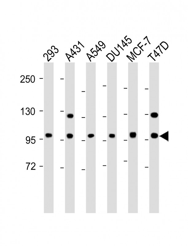
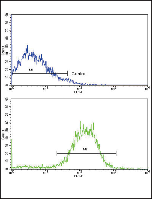
E Cadherin (CDH1) Antibody (N-term)
Purified Rabbit Polyclonal Antibody (Pab)
- SPECIFICATION
- CITATIONS: 8
- PROTOCOLS
- BACKGROUND

Application
| WB, FC, E |
|---|---|
| Primary Accession | P12830 |
| Reactivity | Human |
| Host | Rabbit |
| Clonality | Polyclonal |
| Isotype | Rabbit IgG |
| Calculated MW | 97456 Da |
| Antigen Region | 160-189 aa |
| Gene ID | 999 |
|---|---|
| Other Names | Cadherin-1, CAM 120/80, Epithelial cadherin, E-cadherin, Uvomorulin, CD324, E-Cad/CTF1, E-Cad/CTF2, E-Cad/CTF3, CDH1, CDHE, UVO |
| Target/Specificity | This E Cadherin (CDH1) antibody is generated from rabbits immunized with a KLH conjugated synthetic peptide between 160-189 amino acids from the N-terminal region of human E Cadherin (CDH1). |
| Dilution | WB~~1:2000 FC~~1:10~50 E~~Use at an assay dependent concentration. |
| Format | Purified polyclonal antibody supplied in PBS with 0.09% (W/V) sodium azide. This antibody is prepared by Saturated Ammonium Sulfate (SAS) precipitation followed by dialysis against PBS. |
| Storage | Maintain refrigerated at 2-8°C for up to 2 weeks. For long term storage store at -20°C in small aliquots to prevent freeze-thaw cycles. |
| Precautions | E Cadherin (CDH1) Antibody (N-term) is for research use only and not for use in diagnostic or therapeutic procedures. |
| Name | CDH1 (HGNC:1748) |
|---|---|
| Function | Cadherins are calcium-dependent cell adhesion proteins (PubMed:11976333). They preferentially interact with themselves in a homophilic manner in connecting cells; cadherins may thus contribute to the sorting of heterogeneous cell types. CDH1 is involved in mechanisms regulating cell-cell adhesions, mobility and proliferation of epithelial cells (PubMed:11976333). Promotes organization of radial actin fiber structure and cellular response to contractile forces, via its interaction with AMOTL2 which facilitates anchoring of radial actin fibers to CDH1 junction complexes at the cell membrane (By similarity). Plays a role in the early stages of desmosome cell-cell junction formation via facilitating the recruitment of DSG2 and DSP to desmosome plaques (PubMed:29999492). Has a potent invasive suppressor role. It is a ligand for integrin alpha-E/beta-7. |
| Cellular Location | Cell junction, adherens junction. Cell membrane; Single-pass type I membrane protein. Endosome. Golgi apparatus, trans-Golgi network. Cytoplasm. Cell junction, desmosome. Note=Colocalizes with DLGAP5 at sites of cell-cell contact in intestinal epithelial cells. Anchored to actin microfilaments through association with alpha-, beta- and gamma- catenin. Sequential proteolysis induced by apoptosis or calcium influx, results in translocation from sites of cell-cell contact to the cytoplasm. Colocalizes with RAB11A endosomes during its transport from the Golgi apparatus to the plasma membrane. Recruited to desmosomes at the initial assembly phase and also accumulates progressively at mature desmosome cell-cell junctions (PubMed:25208567, PubMed:29999492) Localizes to cell-cell contacts as keratinocyte differentiation progresses (By similarity). {ECO:0000250|UniProtKB:P09803, ECO:0000269|PubMed:25208567, ECO:0000269|PubMed:29999492} |
| Tissue Location | Expressed in granuloma macrophages (at protein level) (PubMed:27760340). Expressed in the skin (at protein level) (PubMed:22294297). Expressed in the liver (PubMed:3263290) |

Provided below are standard protocols that you may find useful for product applications.
Background
CDH1 is a classical cadherin from the cadherin superfamily. This protein is a calcium dependent cell-cell adhesion glycoprotein comprised of five extracellular cadherin repeats, a transmembrane region and a highly conserved cytoplasmic tail. Mutations are correlated with gastric, breast, colorectal, thyroid and ovarian cancer. Loss of function is thought to contribute to progression in cancer by increasing proliferation, invasion, and/or metastasis. The ectodomain of this protein mediates bacterial adhesion to mammalian cells and the cytoplasmic domain is required for internalization.
References
Mansouri,A., Differentiation 38 (1), 67-71 (1988) Knudsen,K.A. J. Cell Biol. 118 (3), 671-679 (1992) Hsu,Y.M., Cancer Res. 67 (22), 11064-11073 (2007)
If you have used an Abcepta product and would like to share how it has performed, please click on the "Submit Review" button and provide the requested information. Our staff will examine and post your review and contact you if needed.
If you have any additional inquiries please email technical services at tech@abcepta.com.
Ordering Information
Other Products
Shipping Information
