


 Foundational characteristics of cancer include proliferation, angiogenesis, migration, evasion of apoptosis, and cellular immortality. Find key markers for these cellular processes and antibodies to detect them.
 The SUMOplot™ Analysis Program predicts and scores sumoylation sites in your protein. SUMOylation is a post-translational modification involved in various cellular processes, such as nuclear-cytosolic transport, transcriptional regulation, apoptosis, protein stability, response to stress, and progression through the cell cycle.
 The Autophagy Receptor Motif Plotter predicts and scores autophagy receptor binding sites in your protein. Identifying proteins connected to this pathway is critical to understanding the role of autophagy in physiological as well as pathological processes such as development, differentiation, neurodegenerative diseases, stress, infection, and cancer.
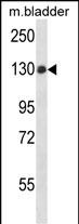
PTPN14 Antibody (C-term)
Affinity Purified Rabbit Polyclonal Antibody (Pab)
- SPECIFICATION
- CITATIONS
- PROTOCOLS
- BACKGROUND

Application
| WB, E |
|---|---|
| Primary Accession | Q15678 |
| Other Accession | NP_005392.2 |
| Reactivity | Mouse |
| Host | Rabbit |
| Clonality | Polyclonal |
| Isotype | Rabbit IgG |
| Calculated MW | 135261 Da |
| Antigen Region | 862-890 aa |
| Gene ID | 5784 |
|---|---|
| Other Names | Tyrosine-protein phosphatase non-receptor type 14, Protein-tyrosine phosphatase pez, PTPN14, PEZ, PTPD2 |
| Target/Specificity | This PTPN14 antibody is generated from rabbits immunized with a KLH conjugated synthetic peptide between 862-890 amino acids from the C-terminal region of human PTPN14. |
| Dilution | WB~~1:1000 E~~Use at an assay dependent concentration. |
| Format | Purified polyclonal antibody supplied in PBS with 0.09% (W/V) sodium azide. This antibody is purified through a protein A column, followed by peptide affinity purification. |
| Storage | Maintain refrigerated at 2-8°C for up to 2 weeks. For long term storage store at -20°C in small aliquots to prevent freeze-thaw cycles. |
| Precautions | PTPN14 Antibody (C-term) is for research use only and not for use in diagnostic or therapeutic procedures. |
| Name | PTPN14 |
|---|---|
| Synonyms | PEZ, PTPD2 |
| Function | Protein tyrosine phosphatase which may play a role in the regulation of lymphangiogenesis, cell-cell adhesion, cell-matrix adhesion, cell migration, cell growth and also regulates TGF-beta gene expression, thereby modulating epithelial-mesenchymal transition. Mediates beta-catenin dephosphorylation at adhesion junctions. Acts as a negative regulator of the oncogenic property of YAP, a downstream target of the hippo pathway, in a cell density-dependent manner. May function as a tumor suppressor. |
| Cellular Location | Cytoplasm. Cytoplasm, cytoskeleton. Nucleus. Note=Translocation into the nucleus is associated with induction of cell proliferation. Partially colocalized with actin filaments at the plasma membrane |
| Tissue Location | Ubiquitous. |

Thousands of laboratories across the world have published research that depended on the performance of antibodies from Abcepta to advance their research. Check out links to articles that cite our products in major peer-reviewed journals, organized by research category.
info@abcepta.com, and receive a free "I Love Antibodies" mug.

Provided below are standard protocols that you may find useful for product applications.
Background
The protein encoded by this gene is a member of the protein tyrosine phosphatase (PTP) family. PTPs are known to be signaling molecules that regulate a variety of cellular processes including cell growth, differentiation, mitotic cycle, and oncogenic transformation. This PTP contains an N-terminal noncatalytic domain similar to that of band 4.1 superfamily cytoskeleton-associated proteins, which suggested the membrane or cytoskeleton localization of this protein. It appears to regulate lymphatic development in mammals, and a loss of function mutation has been found in a kindred with a lymphedema-choanal atresia.
References
Au, A.C., et al. Am. J. Hum. Genet. 87(3):436-444(2010)
Rose, J.E., et al. Mol. Med. 16 (7-8), 247-253 (2010) :
Wyatt, L., et al. Cell Cycle 7(15):2290-2295(2008)
Matsuoka, S., et al. Science 316(5828):1160-1166(2007)
Olsen, J.V., et al. Cell 127(3):635-648(2006)
If you have used an Abcepta product and would like to share how it has performed, please click on the "Submit Review" button and provide the requested information. Our staff will examine and post your review and contact you if needed.
If you have any additional inquiries please email technical services at tech@abcepta.com.
Ordering Information
Other Products
Shipping Information
