


 Foundational characteristics of cancer include proliferation, angiogenesis, migration, evasion of apoptosis, and cellular immortality. Find key markers for these cellular processes and antibodies to detect them.
 The SUMOplot™ Analysis Program predicts and scores sumoylation sites in your protein. SUMOylation is a post-translational modification involved in various cellular processes, such as nuclear-cytosolic transport, transcriptional regulation, apoptosis, protein stability, response to stress, and progression through the cell cycle.
 The Autophagy Receptor Motif Plotter predicts and scores autophagy receptor binding sites in your protein. Identifying proteins connected to this pathway is critical to understanding the role of autophagy in physiological as well as pathological processes such as development, differentiation, neurodegenerative diseases, stress, infection, and cancer.
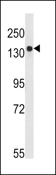
BDP (ARID3B) Antibody (C-term)
Affinity Purified Rabbit Polyclonal Antibody (Pab)
- SPECIFICATION
- CITATIONS
- PROTOCOLS
- BACKGROUND

Application
| WB, IHC-P, E |
|---|---|
| Primary Accession | Q8IVW6 |
| Reactivity | Human |
| Host | Rabbit |
| Clonality | Polyclonal |
| Isotype | Rabbit IgG |
| Calculated MW | 60637 Da |
| Antigen Region | 409-439 aa |
| Gene ID | 10620 |
|---|---|
| Other Names | AT-rich interactive domain-containing protein 3B, ARID domain-containing protein 3B, Bright and dead ringer protein, Bright-like protein, ARID3B, BDP, DRIL2 |
| Target/Specificity | This BDP (ARID3B) antibody is generated from rabbits immunized with a KLH conjugated synthetic peptide between 409-439 amino acids from the C-terminal region of human BDP (ARID3B). |
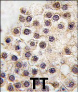
| Dilution | WB~~1:1000 IHC-P~~1:10~50 E~~Use at an assay dependent concentration. |
| Format | Purified polyclonal antibody supplied in PBS with 0.09% (W/V) sodium azide. This antibody is prepared by Saturated Ammonium Sulfate (SAS) precipitation followed by dialysis against PBS. |
| Storage | Maintain refrigerated at 2-8°C for up to 2 weeks. For long term storage store at -20°C in small aliquots to prevent freeze-thaw cycles. |
| Precautions | BDP (ARID3B) Antibody (C-term) is for research use only and not for use in diagnostic or therapeutic procedures. |
| Name | ARID3B |
|---|---|
| Synonyms | BDP, DRIL2 |
| Function | Transcription factor which may be involved in neuroblastoma growth and malignant transformation. Favors nuclear targeting of ARID3A. |
| Cellular Location | Nucleus {ECO:0000255|PROSITE-ProRule:PRU00355, ECO:0000269|PubMed:10446990, ECO:0000269|PubMed:17400556} |
| Tissue Location | Expressed in placenta, testis and leukocytes. Expressed in neuroblastoma. Present in K-562 erythrocytic leukemia cell line (at protein level). |

Thousands of laboratories across the world have published research that depended on the performance of antibodies from Abcepta to advance their research. Check out links to articles that cite our products in major peer-reviewed journals, organized by research category.
info@abcepta.com, and receive a free "I Love Antibodies" mug.

Provided below are standard protocols that you may find useful for product applications.
Background
ARIDB3 is a member of the ARID (AT-rich interaction domain) family of DNA-binding proteins. This protein is homologous with two proteins that bind to the retinoblastoma gene product, and also with the mouse Bright and Drosophila dead ringer proteins. Members of the ARID family have roles in embryonic patterning, cell lineage gene regulation, cell cycle control, transcriptional regulation and possibly in chromatin structure modification.
References
Kortschak,R.D., Trends Biochem. Sci. 25 (6), 294-299 (2000)
Numata,S., Cancer Res. 59 (15), 3741-3747 (1999)
If you have used an Abcepta product and would like to share how it has performed, please click on the "Submit Review" button and provide the requested information. Our staff will examine and post your review and contact you if needed.
If you have any additional inquiries please email technical services at tech@abcepta.com.
Ordering Information
Other Products
Shipping Information
