


 Foundational characteristics of cancer include proliferation, angiogenesis, migration, evasion of apoptosis, and cellular immortality. Find key markers for these cellular processes and antibodies to detect them.
 The SUMOplot™ Analysis Program predicts and scores sumoylation sites in your protein. SUMOylation is a post-translational modification involved in various cellular processes, such as nuclear-cytosolic transport, transcriptional regulation, apoptosis, protein stability, response to stress, and progression through the cell cycle.
 The Autophagy Receptor Motif Plotter predicts and scores autophagy receptor binding sites in your protein. Identifying proteins connected to this pathway is critical to understanding the role of autophagy in physiological as well as pathological processes such as development, differentiation, neurodegenerative diseases, stress, infection, and cancer.
PCSK6 Antibody (C-term)
Affinity Purified Rabbit Polyclonal Antibody (Pab)
- SPECIFICATION
- CITATIONS
- PROTOCOLS
- BACKGROUND

Application
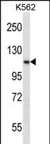
| WB, E |
|---|---|
| Primary Accession | P29122 |
| Other Accession | NP_612197.1, NP_612194.1, NP_612196.1 |
| Reactivity | Human |
| Host | Rabbit |
| Clonality | Polyclonal |
| Isotype | Rabbit IgG |
| Calculated MW | 106420 Da |
| Antigen Region | 659-688 aa |
| Gene ID | 5046 |
|---|---|
| Other Names | Proprotein convertase subtilisin/kexin type 6, 3421-, Paired basic amino acid cleaving enzyme 4, Subtilisin-like proprotein convertase 4, SPC4, Subtilisin/kexin-like protease PACE4, PCSK6, PACE4 |
| Target/Specificity | This PCSK6 antibody is generated from rabbits immunized with a KLH conjugated synthetic peptide between 659-688 amino acids from the C-terminal region of human PCSK6. |
| Dilution | WB~~1:1000 E~~Use at an assay dependent concentration. |
| Format | Purified polyclonal antibody supplied in PBS with 0.09% (W/V) sodium azide. This antibody is purified through a protein A column, followed by peptide affinity purification. |
| Storage | Maintain refrigerated at 2-8°C for up to 2 weeks. For long term storage store at -20°C in small aliquots to prevent freeze-thaw cycles. |
| Precautions | PCSK6 Antibody (C-term) is for research use only and not for use in diagnostic or therapeutic procedures. |
| Name | PCSK6 |
|---|---|
| Synonyms | PACE4 |
| Function | Serine endoprotease that processes various proproteins by cleavage at paired basic amino acids, recognizing the RXXX[KR]R consensus motif. Likely functions in the constitutive secretory pathway, with unique restricted distribution in both neuroendocrine and non-neuroendocrine tissues. |
| Cellular Location | [Isoform PACE4A-I]: Secreted. [Isoform PACE4C]: Endoplasmic reticulum. Note=Not secreted, remains probably in zymogen form in endoplasmic reticulum [Isoform PACE4E-I]: Endomembrane system; Peripheral membrane protein. Note=Retained intracellularly probably through a hydrophobic cluster in their C-terminus [Isoform PACE4B]: Secreted. |
| Tissue Location | Each PACE4 isoform exhibits a unique restricted distribution. Isoform PACE4A-I is expressed in heart, brain, placenta, lung, skeletal muscle, kidney, pancreas, but at comparatively higher levels in the liver. Isoform PACE4A-II is at least expressed in placenta. Isoform PACE4B was only found in the embryonic kidney cell line from which it was isolated. Isoform PACE4C and isoform PACE4D are expressed in placenta. Isoform PACE4E-I is expressed in cerebellum, placenta and pituitary. Isoform PACE4E-II is at least present in cerebellum |

Thousands of laboratories across the world have published research that depended on the performance of antibodies from Abcepta to advance their research. Check out links to articles that cite our products in major peer-reviewed journals, organized by research category.
info@abcepta.com, and receive a free "I Love Antibodies" mug.

Provided below are standard protocols that you may find useful for product applications.
Background
The protein encoded by this gene belongs to the subtilisin-like proprotein convertase family. The members of this family are proprotein convertases that process latent precursor proteins into their biologically active products. This encoded protein is a calcium-dependent serine endoprotease that can cleave precursor protein at their paired basic amino acid processing sites. Some of its substrates are - transforming growth factor beta related proteins, proalbumin, and von Willebrand factor. This gene is thought to play a role in tumor progression. Alternatively spliced transcript variants encoding different isoforms have been identified.
References
Bailey, S.D., et al. Diabetes Care 33(10):2250-2253(2010)
Rose, J.E., et al. Mol. Med. 16 (7-8), 247-253 (2010) :
Fuller, J.A., et al. Invest. Ophthalmol. Vis. Sci. 50(12):5759-5768(2009)
Talmud, P.J., et al. Am. J. Hum. Genet. 85(5):628-642(2009)
Blanchet, M.H., et al. EMBO J. 27(19):2580-2591(2008)
If you have used an Abcepta product and would like to share how it has performed, please click on the "Submit Review" button and provide the requested information. Our staff will examine and post your review and contact you if needed.
If you have any additional inquiries please email technical services at tech@abcepta.com.
Ordering Information
Other Products
Shipping Information
