


 Foundational characteristics of cancer include proliferation, angiogenesis, migration, evasion of apoptosis, and cellular immortality. Find key markers for these cellular processes and antibodies to detect them.
 The SUMOplot™ Analysis Program predicts and scores sumoylation sites in your protein. SUMOylation is a post-translational modification involved in various cellular processes, such as nuclear-cytosolic transport, transcriptional regulation, apoptosis, protein stability, response to stress, and progression through the cell cycle.
 The Autophagy Receptor Motif Plotter predicts and scores autophagy receptor binding sites in your protein. Identifying proteins connected to this pathway is critical to understanding the role of autophagy in physiological as well as pathological processes such as development, differentiation, neurodegenerative diseases, stress, infection, and cancer.
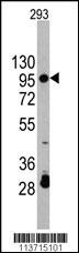
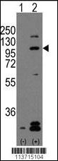
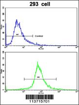
CDH2 Antibody (C-term)
Affinity Purified Rabbit Polyclonal Antibody (Pab)
- SPECIFICATION
- CITATIONS: 1
- PROTOCOLS
- BACKGROUND

Application
| WB, FC, E |
|---|---|
| Primary Accession | P19022 |
| Other Accession | P79883, P39038, P55283, P24503, Q9Z1Y3, P15116, Q90275, P10288, P19534, P20310, P33147 |
| Reactivity | Human |
| Predicted | Xenopus, Bovine, Chicken, Zebrafish, Mouse, Rat |
| Host | Rabbit |
| Clonality | Polyclonal |
| Isotype | Rabbit IgG |
| Calculated MW | 99809 Da |
| Antigen Region | 744-772 aa |
| Gene ID | 1000 |
|---|---|
| Other Names | Cadherin-2, CDw325, Neural cadherin, N-cadherin, CD325, CDH2, CDHN, NCAD |
| Target/Specificity | This CDH2 antibody is generated from rabbits immunized with a KLH conjugated synthetic peptide between 744-772 amino acids from the C-terminal region of human CDH2. |
| Dilution | WB~~1:1000 FC~~1:10~50 E~~Use at an assay dependent concentration. |
| Format | Purified polyclonal antibody supplied in PBS with 0.09% (W/V) sodium azide. This antibody is purified through a protein A column, followed by peptide affinity purification. |
| Storage | Maintain refrigerated at 2-8°C for up to 2 weeks. For long term storage store at -20°C in small aliquots to prevent freeze-thaw cycles. |
| Precautions | CDH2 Antibody (C-term) is for research use only and not for use in diagnostic or therapeutic procedures. |
| Name | CDH2 |
|---|---|
| Synonyms | CDHN, NCAD |
| Function | Calcium-dependent cell adhesion protein; preferentially mediates homotypic cell-cell adhesion by dimerization with a CDH2 chain from another cell. Cadherins may thus contribute to the sorting of heterogeneous cell types. Acts as a regulator of neural stem cells quiescence by mediating anchorage of neural stem cells to ependymocytes in the adult subependymal zone: upon cleavage by MMP24, CDH2-mediated anchorage is affected, leading to modulate neural stem cell quiescence. Plays a role in cell-to-cell junction formation between pancreatic beta cells and neural crest stem (NCS) cells, promoting the formation of processes by NCS cells (By similarity). Required for proper neurite branching. Required for pre- and postsynaptic organization (By similarity). CDH2 may be involved in neuronal recognition mechanism. In hippocampal neurons, may regulate dendritic spine density. |
| Cellular Location | Cell membrane; Single-pass type I membrane protein. Cell membrane, sarcolemma {ECO:0000250|UniProtKB:P15116}. Cell junction. Cell surface {ECO:0000250|UniProtKB:P15116}. Cell junction, desmosome {ECO:0000250|UniProtKB:P15116}. Cell junction, adherens junction {ECO:0000250|UniProtKB:P15116}. Note=Colocalizes with TMEM65 at the intercalated disk in cardiomyocytes. Colocalizes with OBSCN at the intercalated disk and at sarcolemma in cardiomyocytes {ECO:0000250|UniProtKB:P15116} |

Provided below are standard protocols that you may find useful for product applications.
Background
CDH2 is a classical cadherin from the cadherin superfamily. The encoded protein is a calcium dependent cell-cell adhesion glycoprotein comprised of five extracellular cadherin repeats, a transmembrane region and a highly conserved cytoplasmic tail. The protein functions during gastrulation and is required for establishment of left-right asymmetry. At certain central nervous system synapses, presynaptic to postsynaptic adhesion is mediated at least in part by this gene product.
References
Reid R.A., Nucleic Acids Res. 18:5896-5896(1990).
Salomon D., J. Cell Sci. 102:7-17(1992).
Amanchy,R., J. Proteome Res. 4 (5), 1661-1671 (2005)
If you have used an Abcepta product and would like to share how it has performed, please click on the "Submit Review" button and provide the requested information. Our staff will examine and post your review and contact you if needed.
If you have any additional inquiries please email technical services at tech@abcepta.com.
Ordering Information
Other Products
Shipping Information
