


 Foundational characteristics of cancer include proliferation, angiogenesis, migration, evasion of apoptosis, and cellular immortality. Find key markers for these cellular processes and antibodies to detect them.
 The SUMOplot™ Analysis Program predicts and scores sumoylation sites in your protein. SUMOylation is a post-translational modification involved in various cellular processes, such as nuclear-cytosolic transport, transcriptional regulation, apoptosis, protein stability, response to stress, and progression through the cell cycle.
 The Autophagy Receptor Motif Plotter predicts and scores autophagy receptor binding sites in your protein. Identifying proteins connected to this pathway is critical to understanding the role of autophagy in physiological as well as pathological processes such as development, differentiation, neurodegenerative diseases, stress, infection, and cancer.
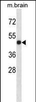
LTBR Antibody (N-term)
Affinity Purified Rabbit Polyclonal Antibody (Pab)
- SPECIFICATION
- CITATIONS
- PROTOCOLS
- BACKGROUND

Application
| WB, E |
|---|---|
| Primary Accession | P36941 |
| Other Accession | NP_002333.1 |
| Reactivity | Mouse |
| Host | Rabbit |
| Clonality | Polyclonal |
| Isotype | Rabbit IgG |
| Calculated MW | 46709 Da |
| Antigen Region | 29-57 aa |
| Gene ID | 4055 |
|---|---|
| Other Names | Tumor necrosis factor receptor superfamily member 3, Lymphotoxin-beta receptor, Tumor necrosis factor C receptor, Tumor necrosis factor receptor 2-related protein, Tumor necrosis factor receptor type III, TNF-RIII, TNFR-III, LTBR, D12S370, TNFCR, TNFR3, TNFRSF3 |
| Target/Specificity | This LTBR antibody is generated from rabbits immunized with a KLH conjugated synthetic peptide between 29-57 amino acids from the N-terminal region of human LTBR. |
| Dilution | WB~~1:1000 E~~Use at an assay dependent concentration. |
| Format | Purified polyclonal antibody supplied in PBS with 0.09% (W/V) sodium azide. This antibody is purified through a protein A column, followed by peptide affinity purification. |
| Storage | Maintain refrigerated at 2-8°C for up to 2 weeks. For long term storage store at -20°C in small aliquots to prevent freeze-thaw cycles. |
| Precautions | LTBR Antibody (N-term) is for research use only and not for use in diagnostic or therapeutic procedures. |
| Name | LTBR |
|---|---|
| Synonyms | D12S370, TNFCR, TNFR3, TNFRSF3 |
| Function | Receptor for the heterotrimeric lymphotoxin containing LTA and LTB, and for TNFS14/LIGHT (PubMed:24248355). Activates NF-kappa-B signaling pathway upon stimulation with lymphotoxin (LTA(1)-LTB(2)) (PubMed:24248355). Promotes apoptosis via TRAF3 and TRAF5. May play a role in the development of lymphoid organs. |
| Cellular Location | Membrane; Single-pass type I membrane protein. |

Thousands of laboratories across the world have published research that depended on the performance of antibodies from Abcepta to advance their research. Check out links to articles that cite our products in major peer-reviewed journals, organized by research category.
info@abcepta.com, and receive a free "I Love Antibodies" mug.

Provided below are standard protocols that you may find useful for product applications.
Background
The protein encoded by this gene is a member of the tumor necrosis factor (TNF) family of receptors. It is expressed on the surface of most cell types, including cells of epithelial and myeloid lineages, but not on T and B lymphocytes. The protein specifically binds the lymphotoxin membrane form (a complex of lymphotoxin-alpha and lymphtoxin-beta). The encoded protein and its ligand play a role in the development and organization of lymphoid tissue and tranformed cells. Activation of the encoded protein can trigger apoptosis.
References
Cheung, T.C., et al. J. Immunol. 185(3):1949-1958(2010)
Han, S., et al. Hum. Immunol. 71(7):727-730(2010)
Sanjo, H., et al. J. Biol. Chem. 285(22):17148-17155(2010)
Rajaraman, P., et al. Cancer Epidemiol. Biomarkers Prev. 19(5):1356-1361(2010)
Davila, S., et al. Genes Immun. 11(3):232-238(2010)
If you have used an Abcepta product and would like to share how it has performed, please click on the "Submit Review" button and provide the requested information. Our staff will examine and post your review and contact you if needed.
If you have any additional inquiries please email technical services at tech@abcepta.com.
Ordering Information
Other Products
Shipping Information
