


 Foundational characteristics of cancer include proliferation, angiogenesis, migration, evasion of apoptosis, and cellular immortality. Find key markers for these cellular processes and antibodies to detect them.
 The SUMOplot™ Analysis Program predicts and scores sumoylation sites in your protein. SUMOylation is a post-translational modification involved in various cellular processes, such as nuclear-cytosolic transport, transcriptional regulation, apoptosis, protein stability, response to stress, and progression through the cell cycle.
 The Autophagy Receptor Motif Plotter predicts and scores autophagy receptor binding sites in your protein. Identifying proteins connected to this pathway is critical to understanding the role of autophagy in physiological as well as pathological processes such as development, differentiation, neurodegenerative diseases, stress, infection, and cancer.
C4BPB Antibody (Center)
Affinity Purified Rabbit Polyclonal Antibody (Pab)
- SPECIFICATION
- CITATIONS
- PROTOCOLS
- BACKGROUND

Application
| WB, E |
|---|---|
| Primary Accession | P20851 |
| Other Accession | NP_001017365.1, NP_001017367.1, NP_000707.1 |
| Reactivity | Human |
| Host | Rabbit |
| Clonality | Polyclonal |
| Isotype | Rabbit IgG |
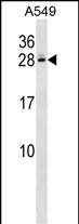
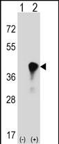
| Calculated MW | 28357 Da |
| Antigen Region | 125-153 aa |
| Gene ID | 725 |
|---|---|
| Other Names | C4b-binding protein beta chain, C4BPB |
| Target/Specificity | This C4BPB antibody is generated from rabbits immunized with a KLH conjugated synthetic peptide between 125-153 amino acids from the Central region of human C4BPB. |
| Dilution | WB~~1:1000 E~~Use at an assay dependent concentration. |
| Format | Purified polyclonal antibody supplied in PBS with 0.09% (W/V) sodium azide. This antibody is purified through a protein A column, followed by peptide affinity purification. |
| Storage | Maintain refrigerated at 2-8°C for up to 2 weeks. For long term storage store at -20°C in small aliquots to prevent freeze-thaw cycles. |
| Precautions | C4BPB Antibody (Center) is for research use only and not for use in diagnostic or therapeutic procedures. |
| Name | C4BPB |
|---|---|
| Function | Controls the classical pathway of complement activation. It binds as a cofactor to C3b/C4b inactivator (C3bINA), which then hydrolyzes the complement fragment C4b. It also accelerates the degradation of the C4bC2a complex (C3 convertase) by dissociating the complement fragment C2a. It also interacts with anticoagulant protein S and with serum amyloid P component. The beta chain binds protein S. |
| Cellular Location | Secreted. |

Thousands of laboratories across the world have published research that depended on the performance of antibodies from Abcepta to advance their research. Check out links to articles that cite our products in major peer-reviewed journals, organized by research category.
info@abcepta.com, and receive a free "I Love Antibodies" mug.

Provided below are standard protocols that you may find useful for product applications.
Background
This gene encodes a member of a superfamily of proteins composed predominantly of tandemly arrayed short consensus repeats of approximately 60 amino acids. A single, unique beta-chain encoded by this gene assembles with seven identical alpha-chains into the predominant isoform of C4b-binding protein, a multimeric protein that controls activation of the complement cascade through the classical pathway. C4b-binding protein has a regulatory role in the coagulation system also, mediated through the beta-chain binding of protein S, a vitamin K-dependent protein that serves as a cofactor of activated protein C. The genes encoding both alpha and beta chains are located adjacent to each other on human chromosome 1 in the regulator of complement activation gene cluster. Alternative splicing gives rise to multiple transcript variants.
References
Han, S., et al. Hum. Immunol. 71(7):727-730(2010)
Rose, J.E., et al. Mol. Med. 16 (7-8), 247-253 (2010) :
Buil, A., et al. Blood 115(23):4644-4650(2010)
Rajaraman, P., et al. Cancer Epidemiol. Biomarkers Prev. 19(5):1356-1361(2010)
Davila, S., et al. Genes Immun. 11(3):232-238(2010)
If you have used an Abcepta product and would like to share how it has performed, please click on the "Submit Review" button and provide the requested information. Our staff will examine and post your review and contact you if needed.
If you have any additional inquiries please email technical services at tech@abcepta.com.
Ordering Information
Other Products
Shipping Information
