


 Foundational characteristics of cancer include proliferation, angiogenesis, migration, evasion of apoptosis, and cellular immortality. Find key markers for these cellular processes and antibodies to detect them.
 The SUMOplot™ Analysis Program predicts and scores sumoylation sites in your protein. SUMOylation is a post-translational modification involved in various cellular processes, such as nuclear-cytosolic transport, transcriptional regulation, apoptosis, protein stability, response to stress, and progression through the cell cycle.
 The Autophagy Receptor Motif Plotter predicts and scores autophagy receptor binding sites in your protein. Identifying proteins connected to this pathway is critical to understanding the role of autophagy in physiological as well as pathological processes such as development, differentiation, neurodegenerative diseases, stress, infection, and cancer.
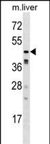
Mouse Camk1g Antibody (C-term)
Affinity Purified Rabbit Polyclonal Antibody (Pab)
- SPECIFICATION
- CITATIONS
- PROTOCOLS
- BACKGROUND

Application
| WB, E |
|---|---|
| Primary Accession | Q91VB2 |
| Other Accession | Q7TNJ7, NP_659066.1 |
| Reactivity | Mouse |
| Predicted | Rat |
| Host | Rabbit |
| Clonality | Polyclonal |
| Isotype | Rabbit IgG |
| Calculated MW | 53296 Da |
| Antigen Region | 426-453 aa |
| Gene ID | 215303 |
|---|---|
| Other Names | Calcium/calmodulin-dependent protein kinase type 1G, CaM kinase I gamma, CaM kinase IG, CaM-KI gamma, CaMKI gamma, CaMKIG, CaMK-like CREB kinase III, CLICK III, Camk1g |
| Target/Specificity | This Mouse Camk1g antibody is generated from rabbits immunized with a KLH conjugated synthetic peptide between 426-453 amino acids from the C-terminal region of mouse Camk1g. |
| Dilution | WB~~1:1000 E~~Use at an assay dependent concentration. |
| Format | Purified polyclonal antibody supplied in PBS with 0.09% (W/V) sodium azide. This antibody is purified through a protein A column, followed by peptide affinity purification. |
| Storage | Maintain refrigerated at 2-8°C for up to 2 weeks. For long term storage store at -20°C in small aliquots to prevent freeze-thaw cycles. |
| Precautions | Mouse Camk1g Antibody (C-term) is for research use only and not for use in diagnostic or therapeutic procedures. |
| Name | Camk1g |
|---|---|
| Function | Calcium/calmodulin-dependent protein kinase belonging to a proposed calcium-triggered signaling cascade. In vitro phosphorylates transcription factor CREB1. |
| Cellular Location | Cytoplasm. Golgi apparatus membrane; Peripheral membrane protein. Cell membrane; Peripheral membrane protein |
| Tissue Location | Highly expressed in brain, in neuronal cell bodies of the central nucleus of amygdala and ventromedial hypothalamic nucleus. Also detected in heart, testis, and kidney |

Thousands of laboratories across the world have published research that depended on the performance of antibodies from Abcepta to advance their research. Check out links to articles that cite our products in major peer-reviewed journals, organized by research category.
info@abcepta.com, and receive a free "I Love Antibodies" mug.

Provided below are standard protocols that you may find useful for product applications.
Background
Calcium/calmodulin-dependent protein kinase belonging to a proposed calcium-triggered signaling cascade. In vitro phosphorylates transcription factor CREB1.
References
Pang, Z.P., et al. J. Biol. Chem. 285(44):33930-33939(2010)
Takemoto-Kimura, S., et al. Neuron 54(5):755-770(2007)
Kamata, A., et al. Neurosci. Res. 57(1):86-97(2007)
Chen, Y., et al. J. Immunol. 175(2):1080-1089(2005)
Frederikse, P.H., et al. Mol. Vis. 10, 794-804 (2004) :
If you have used an Abcepta product and would like to share how it has performed, please click on the "Submit Review" button and provide the requested information. Our staff will examine and post your review and contact you if needed.
If you have any additional inquiries please email technical services at tech@abcepta.com.
Ordering Information
Other Products
Shipping Information
