


 Foundational characteristics of cancer include proliferation, angiogenesis, migration, evasion of apoptosis, and cellular immortality. Find key markers for these cellular processes and antibodies to detect them.
 The SUMOplot™ Analysis Program predicts and scores sumoylation sites in your protein. SUMOylation is a post-translational modification involved in various cellular processes, such as nuclear-cytosolic transport, transcriptional regulation, apoptosis, protein stability, response to stress, and progression through the cell cycle.
 The Autophagy Receptor Motif Plotter predicts and scores autophagy receptor binding sites in your protein. Identifying proteins connected to this pathway is critical to understanding the role of autophagy in physiological as well as pathological processes such as development, differentiation, neurodegenerative diseases, stress, infection, and cancer.
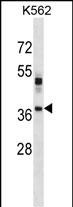
VSX1 Antibody (Center)
Affinity Purified Rabbit Polyclonal Antibody (Pab)
- SPECIFICATION
- CITATIONS
- PROTOCOLS
- BACKGROUND

Application
| WB, E |
|---|---|
| Primary Accession | Q9NZR4 |
| Other Accession | NP_055403.2, NP_955457.1 |
| Reactivity | Human |
| Host | Rabbit |
| Clonality | Polyclonal |
| Isotype | Rabbit IgG |
| Calculated MW | 38431 Da |
| Antigen Region | 125-154 aa |
| Gene ID | 30813 |
|---|---|
| Other Names | Visual system homeobox 1, Homeodomain protein RINX, Retinal inner nuclear layer homeobox protein, Transcription factor VSX1, VSX1, RINX |
| Target/Specificity | This VSX1 antibody is generated from rabbits immunized with a KLH conjugated synthetic peptide between 125-154 amino acids from the Central region of human VSX1. |
| Dilution | WB~~1:1000 E~~Use at an assay dependent concentration. |
| Format | Purified polyclonal antibody supplied in PBS with 0.09% (W/V) sodium azide. This antibody is purified through a protein A column, followed by peptide affinity purification. |
| Storage | Maintain refrigerated at 2-8°C for up to 2 weeks. For long term storage store at -20°C in small aliquots to prevent freeze-thaw cycles. |
| Precautions | VSX1 Antibody (Center) is for research use only and not for use in diagnostic or therapeutic procedures. |
| Name | VSX1 |
|---|---|
| Synonyms | RINX |
| Function | Binds to the 37-bp core of the locus control region (LCR) of the red/green visual pigment gene cluster (PubMed:10903837). May regulate the activity of the LCR and the cone opsin genes at earlier stages of development (PubMed:10903837). Dispensable in early retinal development (By similarity). |
| Cellular Location | Nucleus {ECO:0000250|UniProtKB:Q91V10}. |
| Tissue Location | In the adult eye, expressed in lens, iris, ciliary body, choroid, optical nerve head and, most strongly, in retina, but not expressed in sclera and cornea. According to PubMed:11978762, expressed in adult retina but not in lens and cornea. Within adult retina, found exclusively in the inner nuclear layer. Isoform 1, isoform 2, isoform 3 and isoform 4 expressed in adult retina, but not in brain, heart, kidney, liver, lung, pancreas, placenta and skeletal muscle. Not expressed in thymus and spleen. Expressed in embryonic craniofacial tissue. Expressed in fetal (week 14) retina. Strongly expressed in neonatal retina, weakly in neonatal lens, choroid and cornea (day 1, 4; month 9). |

Thousands of laboratories across the world have published research that depended on the performance of antibodies from Abcepta to advance their research. Check out links to articles that cite our products in major peer-reviewed journals, organized by research category.
info@abcepta.com, and receive a free "I Love Antibodies" mug.

Provided below are standard protocols that you may find useful for product applications.
Background
VSX1 contains a paired-like homeodomain and binds to the core of the locus control region of the red/green visual pigment gene cluster. The encoded protein may regulate expression of the cone opsin genes early in development. Mutations in this gene can cause posterior polymorphous corneal dystrophy and keratoconus.
References
Rose, J.E., et al. Mol. Med. 16 (7-8), 247-253 (2010) :
Dash, D.P., et al. Eye (Lond) 24(6):1085-1092(2010)
Stabuc-Silih, M., et al. Cornea 29(2):172-176(2010)
Stabuc-Silih, M., et al. Acta Dermatovenerol Alp Panonica Adriat 19(2):3-10(2010)
Paliwal, P., et al. Mol. Vis. 15, 2475-2479 (2009) :
If you have used an Abcepta product and would like to share how it has performed, please click on the "Submit Review" button and provide the requested information. Our staff will examine and post your review and contact you if needed.
If you have any additional inquiries please email technical services at tech@abcepta.com.
Ordering Information
Other Products
Shipping Information
