


 Foundational characteristics of cancer include proliferation, angiogenesis, migration, evasion of apoptosis, and cellular immortality. Find key markers for these cellular processes and antibodies to detect them.
 The SUMOplot™ Analysis Program predicts and scores sumoylation sites in your protein. SUMOylation is a post-translational modification involved in various cellular processes, such as nuclear-cytosolic transport, transcriptional regulation, apoptosis, protein stability, response to stress, and progression through the cell cycle.
 The Autophagy Receptor Motif Plotter predicts and scores autophagy receptor binding sites in your protein. Identifying proteins connected to this pathway is critical to understanding the role of autophagy in physiological as well as pathological processes such as development, differentiation, neurodegenerative diseases, stress, infection, and cancer.
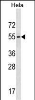
GALR3 Antibody (C-term)
Affinity Purified Rabbit Polyclonal Antibody (Pab)
- SPECIFICATION
- CITATIONS: 1
- PROTOCOLS
- BACKGROUND

Application
| WB, E |
|---|---|
| Primary Accession | O60755 |
| Other Accession | O88626, O88853, NP_003605.1 |
| Reactivity | Human |
| Predicted | Mouse, Rat |
| Host | Rabbit |
| Clonality | Polyclonal |
| Isotype | Rabbit IgG |
| Calculated MW | 39573 Da |
| Antigen Region | 279-307 aa |
| Gene ID | 8484 |
|---|---|
| Other Names | Galanin receptor type 3, GAL3-R, GALR-3, GALR3, GALNR3 |
| Target/Specificity | This GALR3 antibody is generated from rabbits immunized with a KLH conjugated synthetic peptide between 279-307 amino acids from the C-terminal region of human GALR3. |
| Dilution | WB~~1:1000 E~~Use at an assay dependent concentration. |
| Format | Purified polyclonal antibody supplied in PBS with 0.09% (W/V) sodium azide. This antibody is purified through a protein A column, followed by peptide affinity purification. |
| Storage | Maintain refrigerated at 2-8°C for up to 2 weeks. For long term storage store at -20°C in small aliquots to prevent freeze-thaw cycles. |
| Precautions | GALR3 Antibody (C-term) is for research use only and not for use in diagnostic or therapeutic procedures. |
| Name | GALR3 |
|---|---|
| Synonyms | GALNR3 |
| Function | Receptor for the hormone galanin (PubMed:25691535). Receptor for the hormone spexin-1 (PubMed:24517231). |
| Cellular Location | Cell membrane; Multi-pass membrane protein. |

Provided below are standard protocols that you may find useful for product applications.
Background
The neuropeptide galanin modulates a variety of physiologic processes including cognition/memory, sensory/pain processing, hormone secretion, and feeding behavior. The human galanin receptors are G protein-coupled receptors that functionally couple to their intracellular effector through distinct signaling pathways. GALR3 is found in many tissues and may be expressed as 1.4-, 2.4-, and 5-kb transcripts
References
Shimada, M., et al. Hum. Genet. 128(4):433-441(2010)
Saus, E., et al. J Psychiatr Res 44(14):971-978(2010)
Gratacos, M., et al. Am. J. Med. Genet. B Neuropsychiatr. Genet. 150B (6), 808-816 (2009) :
Belfer, I., et al. Genes Brain Behav. 6(5):473-481(2007)
Berger, A., et al. Neuroendocrinology 75(2):130-138(2002)
If you have used an Abcepta product and would like to share how it has performed, please click on the "Submit Review" button and provide the requested information. Our staff will examine and post your review and contact you if needed.
If you have any additional inquiries please email technical services at tech@abcepta.com.
Ordering Information
Other Products
Shipping Information
