


 Foundational characteristics of cancer include proliferation, angiogenesis, migration, evasion of apoptosis, and cellular immortality. Find key markers for these cellular processes and antibodies to detect them.
 The SUMOplot™ Analysis Program predicts and scores sumoylation sites in your protein. SUMOylation is a post-translational modification involved in various cellular processes, such as nuclear-cytosolic transport, transcriptional regulation, apoptosis, protein stability, response to stress, and progression through the cell cycle.
 The Autophagy Receptor Motif Plotter predicts and scores autophagy receptor binding sites in your protein. Identifying proteins connected to this pathway is critical to understanding the role of autophagy in physiological as well as pathological processes such as development, differentiation, neurodegenerative diseases, stress, infection, and cancer.
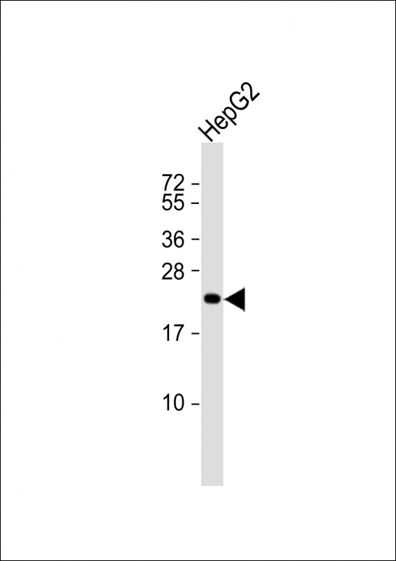
ARL6IP1 Antibody (N-term)
Affinity Purified Rabbit Polyclonal Antibody (Pab)
- SPECIFICATION
- CITATIONS: 2
- PROTOCOLS
- BACKGROUND

Application
| WB, E |
|---|---|
| Primary Accession | Q15041 |
| Other Accession | NP_055976.1 |
| Reactivity | Human |
| Host | Rabbit |
| Clonality | Polyclonal |
| Isotype | Rabbit IgG |
| Calculated MW | 23363 Da |
| Antigen Region | 1-30 aa |
| Gene ID | 23204 |
|---|---|
| Other Names | ADP-ribosylation factor-like protein 6-interacting protein 1, ARL-6-interacting protein 1, Aip-1, ARL6IP1, ARL6IP, KIAA0069 |
| Target/Specificity | This ARL6IP1 antibody is generated from rabbits immunized with a KLH conjugated synthetic peptide between 1-30 amino acids from the N-terminal region of human ARL6IP1. |
| Dilution | WB~~1:1000 E~~Use at an assay dependent concentration. |
| Format | Purified polyclonal antibody supplied in PBS with 0.09% (W/V) sodium azide. This antibody is purified through a protein A column, followed by peptide affinity purification. |
| Storage | Maintain refrigerated at 2-8°C for up to 2 weeks. For long term storage store at -20°C in small aliquots to prevent freeze-thaw cycles. |
| Precautions | ARL6IP1 Antibody (N-term) is for research use only and not for use in diagnostic or therapeutic procedures. |
| Name | ARL6IP1 |
|---|---|
| Function | Positively regulates SLC1A1/EAAC1-mediated glutamate transport by increasing its affinity for glutamate in a PKC activity- dependent manner. Promotes the catalytic efficiency of SLC1A1/EAAC1 probably by reducing its interaction with ARL6IP5, a negative regulator of SLC1A1/EAAC1-mediated glutamate transport (By similarity). Plays a role in the formation and stabilization of endoplasmic reticulum tubules (PubMed:24262037). Negatively regulates apoptosis, possibly by modulating the activity of caspase-9 (CASP9). Inhibits cleavage of CASP9-dependent substrates and downstream markers of apoptosis but not CASP9 itself (PubMed:12754298). May be involved in protein transport, membrane trafficking, or cell signaling during hematopoietic maturation (PubMed:10995579). |
| Cellular Location | Endomembrane system; Multi-pass membrane protein. Endoplasmic reticulum membrane; Multi-pass membrane protein. Endoplasmic reticulum {ECO:0000250|UniProtKB:Q9JKW0}. Note=Predominantly localized to intracytoplasmic membranes. Preferentially localizes at the ER tubules and the edge of the ER sheets, both of which are characterized by a high membrane curvature. |
| Tissue Location | Expressed in all hematopoietic cell lineages, but the highest level of expression is found in early myeloid progenitor cells. Expressed in brain, bone marrow, thymus and lung. Expressed at low level in liver, kidney and spleen. Not detected in heart |

Provided below are standard protocols that you may find useful for product applications.
Background
ARL6IP1 may be involved in protein transport, membrane trafficking, or cell signaling during hematopoietic maturation.
References
Guo, F., et al. Oncol. Rep. 23(5):1449-1455(2010)
Venkatesan, K., et al. Nat. Methods 6(1):83-90(2009)
Lamesch, P., et al. Genomics 89(3):307-315(2007)
Lui, H.M., et al. Mol. Cancer Res. 1(7):508-518(2003)
Pettersson, M., et al. Genomics 68(3):351-354(2000)
If you have used an Abcepta product and would like to share how it has performed, please click on the "Submit Review" button and provide the requested information. Our staff will examine and post your review and contact you if needed.
If you have any additional inquiries please email technical services at tech@abcepta.com.
Ordering Information
Other Products
Shipping Information
