


 Foundational characteristics of cancer include proliferation, angiogenesis, migration, evasion of apoptosis, and cellular immortality. Find key markers for these cellular processes and antibodies to detect them.
 The SUMOplot™ Analysis Program predicts and scores sumoylation sites in your protein. SUMOylation is a post-translational modification involved in various cellular processes, such as nuclear-cytosolic transport, transcriptional regulation, apoptosis, protein stability, response to stress, and progression through the cell cycle.
 The Autophagy Receptor Motif Plotter predicts and scores autophagy receptor binding sites in your protein. Identifying proteins connected to this pathway is critical to understanding the role of autophagy in physiological as well as pathological processes such as development, differentiation, neurodegenerative diseases, stress, infection, and cancer.
EPOR Antibody (Center)
Affinity Purified Rabbit Polyclonal Antibody (Pab)

- SPECIFICATION
- CITATIONS
- PROTOCOLS
- BACKGROUND

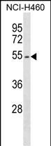
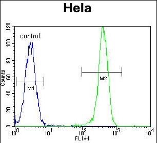
Application
| WB, IF, FC, E |
|---|---|
| Primary Accession | P19235 |
| Other Accession | NP_000112.1 |
| Reactivity | Human |
| Host | Rabbit |
| Clonality | Polyclonal |
| Isotype | Rabbit IgG |
| Calculated MW | 55065 Da |
| Antigen Region | 329-357 aa |
| Gene ID | 2057 |
|---|---|
| Other Names | Erythropoietin receptor, EPO-R, EPOR |
| Target/Specificity | This EPOR antibody is generated from rabbits immunized with a KLH conjugated synthetic peptide between 329-357 amino acids from the Central region of human EPOR. |
| Dilution | WB~~1:1000 IF~~1:10~50 FC~~1:10~50 E~~Use at an assay dependent concentration. |
| Format | Purified polyclonal antibody supplied in PBS with 0.09% (W/V) sodium azide. This antibody is purified through a protein A column, followed by peptide affinity purification. |
| Storage | Maintain refrigerated at 2-8°C for up to 2 weeks. For long term storage store at -20°C in small aliquots to prevent freeze-thaw cycles. |
| Precautions | EPOR Antibody (Center) is for research use only and not for use in diagnostic or therapeutic procedures. |
| Name | EPOR {ECO:0000303|PubMed:2163695, ECO:0000312|HGNC:HGNC:3416} |
|---|---|
| Function | Receptor for erythropoietin, which mediates erythropoietin- induced erythroblast proliferation and differentiation (PubMed:10388848, PubMed:2163695, PubMed:2163696, PubMed:8662939, PubMed:9774108). Upon EPO stimulation, EPOR dimerizes triggering the JAK2/STAT5 signaling cascade (By similarity). In some cell types, can also activate STAT1 and STAT3 (PubMed:11756159). May also activate the LYN tyrosine kinase (By similarity). |
| Cellular Location | Cell membrane {ECO:0000250|UniProtKB:P14753}; Single-pass type I membrane protein |
| Tissue Location | Erythroid cells and erythroid progenitor cells. [Isoform EPOR-S]: Isoform EPOR-S and isoform EPOR-T are the predominant forms in bone marrow. |

Thousands of laboratories across the world have published research that depended on the performance of antibodies from Abcepta to advance their research. Check out links to articles that cite our products in major peer-reviewed journals, organized by research category.
info@abcepta.com, and receive a free "I Love Antibodies" mug.

Provided below are standard protocols that you may find useful for product applications.
Background
This gene encodes the erythropoietin receptor which is a member of the cytokine receptor family. Upon erythropoietin binding, this receptor activates Jak2 tyrosine kinase which activates different intracellular pathways including: Ras/MAP kinase, phosphatidylinositol 3-kinase and STAT transcription factors. The stimulated erythropoietin receptor appears to have a role in erythroid cell survival. Defects in the erythropoietin receptor may produce erythroleukemia and familial erythrocytosis. Dysregulation of this gene may affect the growth of certain tumors. Alternate splicing results in multiple transcript variants.
References
Lim, A.C., et al. Biochemistry 49(18):3797-3804(2010)
Perrotta, S., et al. PLoS ONE 5 (8), E12015 (2010) :
Khankin, E.V., et al. PLoS ONE 5 (2), E9246 (2010) :
Wincewicz, A., et al. Folia Histochem. Cytobiol. 47(3):425-430(2009)
Ketteler, R., et al. J. Biol. Chem. 278(4):2654-2660(2003)
If you have used an Abcepta product and would like to share how it has performed, please click on the "Submit Review" button and provide the requested information. Our staff will examine and post your review and contact you if needed.
If you have any additional inquiries please email technical services at tech@abcepta.com.
Ordering Information
Other Products
Shipping Information
