


 Foundational characteristics of cancer include proliferation, angiogenesis, migration, evasion of apoptosis, and cellular immortality. Find key markers for these cellular processes and antibodies to detect them.
 The SUMOplot™ Analysis Program predicts and scores sumoylation sites in your protein. SUMOylation is a post-translational modification involved in various cellular processes, such as nuclear-cytosolic transport, transcriptional regulation, apoptosis, protein stability, response to stress, and progression through the cell cycle.
 The Autophagy Receptor Motif Plotter predicts and scores autophagy receptor binding sites in your protein. Identifying proteins connected to this pathway is critical to understanding the role of autophagy in physiological as well as pathological processes such as development, differentiation, neurodegenerative diseases, stress, infection, and cancer.
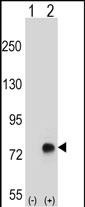
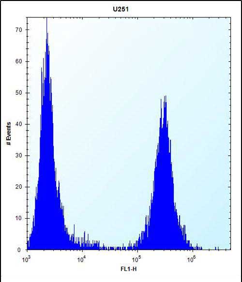
TGFBI Antibody (N-term)
Affinity Purified Rabbit Polyclonal Antibody (Pab)
- SPECIFICATION
- CITATIONS
- PROTOCOLS
- BACKGROUND

Application
| WB, FC, E |
|---|---|
| Primary Accession | Q15582 |
| Other Accession | Q95215, O11780, P82198, NP_000349.1, P55906 |
| Reactivity | Human |
| Predicted | Bovine, Mouse, Pig, Rabbit |
| Host | Rabbit |
| Clonality | Polyclonal |
| Isotype | Rabbit IgG |
| Calculated MW | 74681 Da |
| Antigen Region | 106-135 aa |
| Gene ID | 7045 |
|---|---|
| Other Names | Transforming growth factor-beta-induced protein ig-h3, Beta ig-h3, Kerato-epithelin, RGD-containing collagen-associated protein, RGD-CAP, TGFBI, BIGH3 |
| Target/Specificity | This TGFBI antibody is generated from rabbits immunized with a KLH conjugated synthetic peptide between 106-135 amino acids from the N-terminal region of human TGFBI. |
| Dilution | WB~~1:1000 FC~~1:10~50 E~~Use at an assay dependent concentration. |
| Format | Purified polyclonal antibody supplied in PBS with 0.09% (W/V) sodium azide. This antibody is purified through a protein A column, followed by peptide affinity purification. |
| Storage | Maintain refrigerated at 2-8°C for up to 2 weeks. For long term storage store at -20°C in small aliquots to prevent freeze-thaw cycles. |
| Precautions | TGFBI Antibody (N-term) is for research use only and not for use in diagnostic or therapeutic procedures. |
| Name | TGFBI |
|---|---|
| Synonyms | BIGH3 |
| Function | Plays a role in cell adhesion (PubMed:8024701). May play a role in cell-collagen interactions (By similarity). |
| Cellular Location | Secreted. Secreted, extracellular space, extracellular matrix Note=May be associated both with microfibrils and with the cell surface (PubMed:8077289). |
| Tissue Location | Highly expressed in the corneal epithelium (PubMed:27609313, PubMed:8077289). Expressed in heart, placenta, lung, liver, skeletal muscle, kidney and pancreas (PubMed:8077289) |

Thousands of laboratories across the world have published research that depended on the performance of antibodies from Abcepta to advance their research. Check out links to articles that cite our products in major peer-reviewed journals, organized by research category.
info@abcepta.com, and receive a free "I Love Antibodies" mug.

Provided below are standard protocols that you may find useful for product applications.
Background
This gene encodes an RGD-containing protein that binds to type I, II and IV collagens. The RGD motif is found in many extracellular matrix proteins modulating cell adhesion and serves as a ligand recognition sequence for several integrins. This protein plays a role in cell-collagen interactions and may be involved in endochondrial bone formation in cartilage. The protein is induced by transforming growth factor-beta and acts to inhibit cell adhesion. Mutations in this gene are associated with multiple types of corneal dystrophy.
References
Bailey, S.D., et al. Diabetes Care 33(10):2250-2253(2010)
Edelstein, S.L., et al. Cornea 29(6):698-700(2010)
Romero, P., et al. Mol. Vis. 16, 1601-1609 (2010) :
Paliwal, P., et al. Mol. Vis. 16, 1429-1438 (2010) :
Yang, J., et al. Mol. Vis. 16, 1186-1193 (2010) :
If you have used an Abcepta product and would like to share how it has performed, please click on the "Submit Review" button and provide the requested information. Our staff will examine and post your review and contact you if needed.
If you have any additional inquiries please email technical services at tech@abcepta.com.
Ordering Information
Other Products
Shipping Information
