


 Foundational characteristics of cancer include proliferation, angiogenesis, migration, evasion of apoptosis, and cellular immortality. Find key markers for these cellular processes and antibodies to detect them.
 The SUMOplot™ Analysis Program predicts and scores sumoylation sites in your protein. SUMOylation is a post-translational modification involved in various cellular processes, such as nuclear-cytosolic transport, transcriptional regulation, apoptosis, protein stability, response to stress, and progression through the cell cycle.
 The Autophagy Receptor Motif Plotter predicts and scores autophagy receptor binding sites in your protein. Identifying proteins connected to this pathway is critical to understanding the role of autophagy in physiological as well as pathological processes such as development, differentiation, neurodegenerative diseases, stress, infection, and cancer.
SLC2A9 Antibody (N-term)
Affinity Purified Rabbit Polyclonal Antibody (Pab)
- SPECIFICATION
- CITATIONS
- PROTOCOLS
- BACKGROUND

Application
| WB, E |
|---|---|
| Primary Accession | Q9NRM0 |
| Other Accession | NP_064425.2, NP_001001290.1 |
| Reactivity | Human |
| Host | Rabbit |
| Clonality | Polyclonal |
| Isotype | Rabbit IgG |
| Calculated MW | 58702 Da |
| Antigen Region | 75-104 aa |
| Gene ID | 56606 |
|---|---|
| Other Names | Solute carrier family 2, facilitated glucose transporter member 9, Glucose transporter type 9, GLUT-9, SLC2A9, GLUT9 |
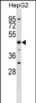
| Target/Specificity | This SLC2A9 antibody is generated from rabbits immunized with a KLH conjugated synthetic peptide between 75-104 amino acids from the N-terminal region of human SLC2A9. |
| Dilution | WB~~1:1000 E~~Use at an assay dependent concentration. |
| Format | Purified polyclonal antibody supplied in PBS with 0.09% (W/V) sodium azide. This antibody is purified through a protein A column, followed by peptide affinity purification. |
| Storage | Maintain refrigerated at 2-8°C for up to 2 weeks. For long term storage store at -20°C in small aliquots to prevent freeze-thaw cycles. |
| Precautions | SLC2A9 Antibody (N-term) is for research use only and not for use in diagnostic or therapeutic procedures. |
| Name | SLC2A9 {ECO:0000303|PubMed:10860667, ECO:0000312|HGNC:HGNC:13446} |
|---|---|
| Function | High-capacity urate transporter, which may play a role in the urate reabsorption by proximal tubules (PubMed:18327257, PubMed:18701466, PubMed:22647630, PubMed:28083649, PubMed:36749388). May have a residual high-affinity, low-capacity glucose and fructose transporter activity (PubMed:18327257, PubMed:18701466, PubMed:18842065). Transports urate at rates 45- to 60-fold faster than glucose (PubMed:18842065). Does not transport galactose (PubMed:28083649). May mediate small uptake of adenine but not of other nucleobases (PubMed:22647630). |
| Cellular Location | [Isoform 1]: Cell membrane; Multi-pass membrane protein. Basolateral cell membrane; Multi-pass membrane protein |
| Tissue Location | [Isoform 1]: Most strongly expressed in basolateral membranes of proximal renal tubular cells, liver and placenta. Also detected in lung, blood leukocytes, heart skeletal muscle and chondrocytes from articular cartilage. Detected in kidney membrane (at protein level). |

Thousands of laboratories across the world have published research that depended on the performance of antibodies from Abcepta to advance their research. Check out links to articles that cite our products in major peer-reviewed journals, organized by research category.
info@abcepta.com, and receive a free "I Love Antibodies" mug.

Provided below are standard protocols that you may find useful for product applications.
Background
This gene encodes a member of the SLC2A facilitative glucose transporter family. Members of this family play a significant role in maintaining glucose homeostasis. The encoded protein may play a role in the development and survival of chondrocytes in cartilage matrices. Two transcript variants encoding distinct isoforms have been identified for this gene.
References
Rose, J.E., et al. Mol. Med. 16 (7-8), 247-253 (2010) :
Facheris, M.F., et al. J. Mol. Neurosci. (2010) In press :
Houlihan, L.M., et al. Hum. Mol. Genet. 19(11):2321-2330(2010)
Urano, W., et al. Ann. Rheum. Dis. 69(5):932-933(2010)
Tabara, Y., et al. Am. J. Nephrol. 32(3):279-286(2010)
If you have used an Abcepta product and would like to share how it has performed, please click on the "Submit Review" button and provide the requested information. Our staff will examine and post your review and contact you if needed.
If you have any additional inquiries please email technical services at tech@abcepta.com.
Ordering Information
Shipping Information
