


 Foundational characteristics of cancer include proliferation, angiogenesis, migration, evasion of apoptosis, and cellular immortality. Find key markers for these cellular processes and antibodies to detect them.
 The SUMOplot™ Analysis Program predicts and scores sumoylation sites in your protein. SUMOylation is a post-translational modification involved in various cellular processes, such as nuclear-cytosolic transport, transcriptional regulation, apoptosis, protein stability, response to stress, and progression through the cell cycle.
 The Autophagy Receptor Motif Plotter predicts and scores autophagy receptor binding sites in your protein. Identifying proteins connected to this pathway is critical to understanding the role of autophagy in physiological as well as pathological processes such as development, differentiation, neurodegenerative diseases, stress, infection, and cancer.
PRKAG3 Antibody (C-term)
Affinity Purified Rabbit Polyclonal Antibody (Pab)
- SPECIFICATION
- CITATIONS
- PROTOCOLS
- BACKGROUND

Application
| WB, E |
|---|---|
| Primary Accession | Q9UGI9 |
| Other Accession | Q8BGM7, Q2LL38, NP_059127.2 |
| Reactivity | Human |
| Predicted | Bovine, Mouse |
| Host | Rabbit |
| Clonality | Polyclonal |
| Isotype | Rabbit IgG |
| Calculated MW | 54258 Da |
| Antigen Region | 426-454 aa |
| Gene ID | 53632 |
|---|---|
| Other Names | 5'-AMP-activated protein kinase subunit gamma-3, AMPK gamma3, AMPK subunit gamma-3, PRKAG3, AMPKG3 |
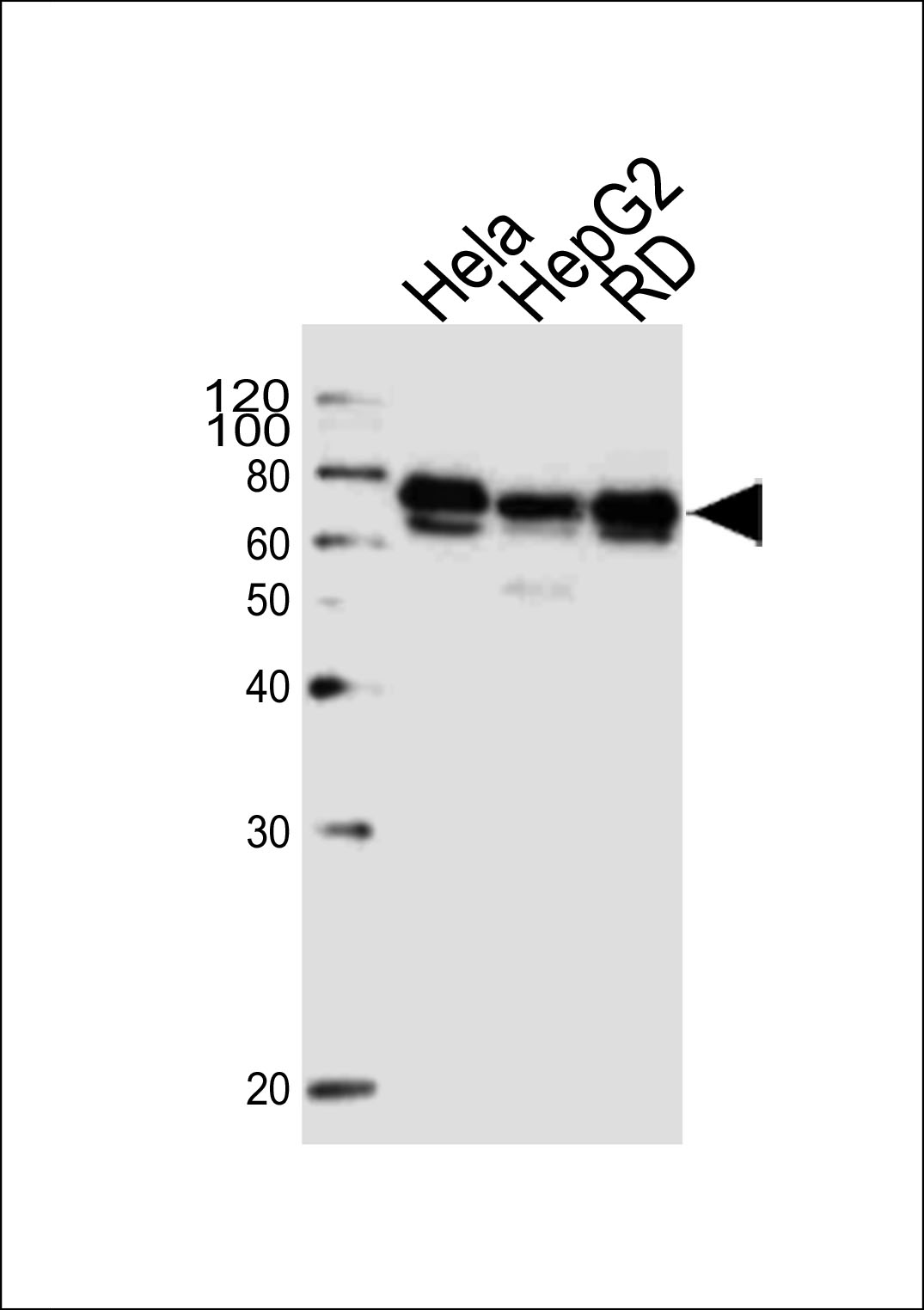
| Target/Specificity | This PRKAG3 antibody is generated from rabbits immunized with a KLH conjugated synthetic peptide between 426-454 amino acids from the C-terminal region of human PRKAG3. |
| Dilution | WB~~1:1000 E~~Use at an assay dependent concentration. |
| Format | Purified polyclonal antibody supplied in PBS with 0.09% (W/V) sodium azide. This antibody is purified through a protein A column, followed by peptide affinity purification. |
| Storage | Maintain refrigerated at 2-8°C for up to 2 weeks. For long term storage store at -20°C in small aliquots to prevent freeze-thaw cycles. |
| Precautions | PRKAG3 Antibody (C-term) is for research use only and not for use in diagnostic or therapeutic procedures. |
| Name | PRKAG3 |
|---|---|
| Synonyms | AMPKG3 |
| Function | AMP/ATP-binding subunit of AMP-activated protein kinase (AMPK), an energy sensor protein kinase that plays a key role in regulating cellular energy metabolism (PubMed:14722619, PubMed:17878938, PubMed:24563466). In response to reduction of intracellular ATP levels, AMPK activates energy-producing pathways and inhibits energy-consuming processes: inhibits protein, carbohydrate and lipid biosynthesis, as well as cell growth and proliferation. AMPK acts via direct phosphorylation of metabolic enzymes, and by longer-term effects via phosphorylation of transcription regulators. AMPK also acts as a regulator of cellular polarity by remodeling the actin cytoskeleton; probably by indirectly activating myosin. The AMPK gamma3 subunit is a non-catalytic subunit with a regulatory role in muscle energy metabolism (PubMed:17878938). It mediates binding to AMP, ADP and ATP, leading to AMPK activation or inhibition: AMP-binding results in allosteric activation of alpha catalytic subunit (PRKAA1 or PRKAA2) both by inducing phosphorylation and preventing dephosphorylation of catalytic subunits. ADP also stimulates phosphorylation, without stimulating already phosphorylated catalytic subunit. ATP promotes dephosphorylation of catalytic subunit, rendering the AMPK enzyme inactive. |
| Tissue Location | Skeletal muscle, with weak expression in heart and pancreas |

Thousands of laboratories across the world have published research that depended on the performance of antibodies from Abcepta to advance their research. Check out links to articles that cite our products in major peer-reviewed journals, organized by research category.
info@abcepta.com, and receive a free "I Love Antibodies" mug.

Provided below are standard protocols that you may find useful for product applications.
Background
The protein encoded by this gene is a regulatory subunit of the AMP-activated protein kinase (AMPK). AMPK is a heterotrimer consisting of an alpha catalytic subunit, and non-catalytic beta and gamma subunits. AMPK is an important energy-sensing enzyme that monitors cellular energy status. In response to cellular metabolic stresses, AMPK is activated, and thus phosphorylates and inactivates acetyl-CoA carboxylase (ACC) and beta-hydroxy beta-methylglutaryl-CoA reductase (HMGCR), key enzymes involved in regulating de novo biosynthesis of fatty acid and cholesterol. This subunit is one of the gamma regulatory subunits of AMPK. It is dominantly expressed in skeletal muscle. Studies of the pig counterpart suggest that this subunit may play a key role in the regulation of energy metabolism in skeletal muscle. [provided by RefSeq].
References
Jablonski, K.A., et al. Diabetes 59(10):2672-2681(2010)
Jassim, G., et al. Pharmacopsychiatry (2010) In press :
Crawford, S.A., et al. Diabetologia 53(9):1986-1997(2010)
Ramanathan, L., et al. Protein Expr. Purif. 70(1):13-22(2010)
McGeachie, M., et al. Circulation 120(24):2448-2454(2009)
If you have used an Abcepta product and would like to share how it has performed, please click on the "Submit Review" button and provide the requested information. Our staff will examine and post your review and contact you if needed.
If you have any additional inquiries please email technical services at tech@abcepta.com.
Ordering Information
Other Products
Shipping Information
