


 Foundational characteristics of cancer include proliferation, angiogenesis, migration, evasion of apoptosis, and cellular immortality. Find key markers for these cellular processes and antibodies to detect them.
 The SUMOplot™ Analysis Program predicts and scores sumoylation sites in your protein. SUMOylation is a post-translational modification involved in various cellular processes, such as nuclear-cytosolic transport, transcriptional regulation, apoptosis, protein stability, response to stress, and progression through the cell cycle.
 The Autophagy Receptor Motif Plotter predicts and scores autophagy receptor binding sites in your protein. Identifying proteins connected to this pathway is critical to understanding the role of autophagy in physiological as well as pathological processes such as development, differentiation, neurodegenerative diseases, stress, infection, and cancer.
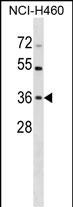
HUS1 Antibody (C-term)
Affinity Purified Rabbit Polyclonal Antibody (Pab)
- SPECIFICATION
- CITATIONS
- PROTOCOLS
- BACKGROUND

Application
| WB, E |
|---|---|
| Primary Accession | O60921 |
| Other Accession | NP_004498.1 |
| Reactivity | Human |
| Host | Rabbit |
| Clonality | Polyclonal |
| Isotype | Rabbit IgG |
| Calculated MW | 31691 Da |
| Antigen Region | 199-226 aa |
| Gene ID | 3364 |
|---|---|
| Other Names | Checkpoint protein HUS1, hHUS1, HUS1 |
| Target/Specificity | This HUS1 antibody is generated from rabbits immunized with a KLH conjugated synthetic peptide between 199-226 amino acids from the C-terminal region of human HUS1. |
| Dilution | WB~~1:1000 E~~Use at an assay dependent concentration. |
| Format | Purified polyclonal antibody supplied in PBS with 0.09% (W/V) sodium azide. This antibody is purified through a protein A column, followed by peptide affinity purification. |
| Storage | Maintain refrigerated at 2-8°C for up to 2 weeks. For long term storage store at -20°C in small aliquots to prevent freeze-thaw cycles. |
| Precautions | HUS1 Antibody (C-term) is for research use only and not for use in diagnostic or therapeutic procedures. |
| Name | HUS1 |
|---|---|
| Function | Component of the 9-1-1 cell-cycle checkpoint response complex that plays a major role in DNA repair (PubMed:21659603). The 9-1-1 complex is recruited to DNA lesion upon damage by the RAD17-replication factor C (RFC) clamp loader complex (PubMed:21659603). Acts then as a sliding clamp platform on DNA for several proteins involved in long- patch base excision repair (LP-BER) (PubMed:21659603). The 9-1-1 complex stimulates DNA polymerase beta (POLB) activity by increasing its affinity for the 3'-OH end of the primer-template and stabilizes POLB to those sites where LP-BER proceeds; endonuclease FEN1 cleavage activity on substrates with double, nick, or gap flaps of distinct sequences and lengths; and DNA ligase I (LIG1) on long-patch base excision repair substrates (PubMed:21659603). The 9-1-1 complex is necessary for the recruitment of RHNO1 to sites of double-stranded breaks (DSB) occurring during the S phase (PubMed:21659603). |
| Cellular Location | Nucleus. Cytoplasm, cytosol. Note=In discrete nuclear foci upon DNA damage (PubMed:11077446). According to PubMed:11077446, localized also in the cytoplasm (PubMed:11077446). DNA damage induces its nuclear translocation (PubMed:11077446). Shuttles between the nucleus and the cytoplasm (PubMed:11077446). |
| Tissue Location | Ubiquitous.. |

Thousands of laboratories across the world have published research that depended on the performance of antibodies from Abcepta to advance their research. Check out links to articles that cite our products in major peer-reviewed journals, organized by research category.
info@abcepta.com, and receive a free "I Love Antibodies" mug.

Provided below are standard protocols that you may find useful for product applications.
Background
The protein encoded by this gene is a component of an evolutionarily conserved, genotoxin-activated checkpoint complex that is involved in the cell cycle arrest in response to DNA damage. This protein forms a heterotrimeric complex with checkpoint proteins RAD9 and RAD1. In response to DNA damage, the trimeric complex interacts with another protein complex consisting of checkpoint protein RAD17 and four small subunits of the replication factor C (RFC), which loads the combined complex onto the chromatin. The DNA damage induced chromatin binding has been shown to depend on the activation of the checkpoint kinase ATM, and is thought to be an early checkpoint signaling event. [provided by RefSeq].
References
Liu, C.Y., et al. Carcinogenesis 31(7):1259-1263(2010)
Takeishi, Y., et al. Genes Cells 15(7):761-771(2010)
Bai, H., et al. DNA Repair (Amst.) 9(5):478-487(2010)
Guey, L.T., et al. Eur. Urol. 57(2):283-292(2010)
Hosgood, H.D. III, et al. Respir Med 103(12):1866-1870(2009)
If you have used an Abcepta product and would like to share how it has performed, please click on the "Submit Review" button and provide the requested information. Our staff will examine and post your review and contact you if needed.
If you have any additional inquiries please email technical services at tech@abcepta.com.
Ordering Information
Shipping Information
