


 Foundational characteristics of cancer include proliferation, angiogenesis, migration, evasion of apoptosis, and cellular immortality. Find key markers for these cellular processes and antibodies to detect them.
 The SUMOplot™ Analysis Program predicts and scores sumoylation sites in your protein. SUMOylation is a post-translational modification involved in various cellular processes, such as nuclear-cytosolic transport, transcriptional regulation, apoptosis, protein stability, response to stress, and progression through the cell cycle.
 The Autophagy Receptor Motif Plotter predicts and scores autophagy receptor binding sites in your protein. Identifying proteins connected to this pathway is critical to understanding the role of autophagy in physiological as well as pathological processes such as development, differentiation, neurodegenerative diseases, stress, infection, and cancer.
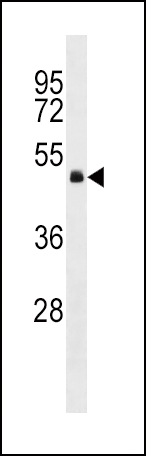
GNA13 Antibody (Center)
Affinity Purified Rabbit Polyclonal Antibody (Pab)
- SPECIFICATION
- CITATIONS
- PROTOCOLS
- BACKGROUND

Application
| WB, E |
|---|---|
| Primary Accession | Q14344 |
| Other Accession | Q6Q7Y5, P27601, NP_006563.2 |
| Reactivity | Human |
| Predicted | Mouse, Rat |
| Host | Rabbit |
| Clonality | Polyclonal |
| Isotype | Rabbit IgG |
| Calculated MW | 44050 Da |
| Antigen Region | 184-212 aa |
| Gene ID | 10672 |
|---|---|
| Other Names | Guanine nucleotide-binding protein subunit alpha-13, G alpha-13, G-protein subunit alpha-13, GNA13 |
| Target/Specificity | This GNA13 antibody is generated from rabbits immunized with a KLH conjugated synthetic peptide between 184-212 amino acids from the Central region of human GNA13. |
| Dilution | WB~~1:1000 E~~Use at an assay dependent concentration. |
| Format | Purified polyclonal antibody supplied in PBS with 0.09% (W/V) sodium azide. This antibody is purified through a protein A column, followed by peptide affinity purification. |
| Storage | Maintain refrigerated at 2-8°C for up to 2 weeks. For long term storage store at -20°C in small aliquots to prevent freeze-thaw cycles. |
| Precautions | GNA13 Antibody (Center) is for research use only and not for use in diagnostic or therapeutic procedures. |
| Name | GNA13 |
|---|---|
| Function | Guanine nucleotide-binding proteins (G proteins) are involved as modulators or transducers in various transmembrane signaling systems (PubMed:15240885, PubMed:16705036, PubMed:16787920, PubMed:27084452). Activates effector molecule RhoA by binding and activating RhoGEFs (ARHGEF1/p115RhoGEF, ARHGEF11/PDZ-RhoGEF and ARHGEF12/LARG) (PubMed:12515866, PubMed:15240885). GNA13-dependent Rho signaling subsequently regulates transcription factor AP-1 (activating protein-1) (By similarity). Promotes tumor cell invasion and metastasis by activating RhoA/ROCK signaling pathway (PubMed:16705036, PubMed:16787920, PubMed:27084452). Inhibits CDH1-mediated cell adhesion in a process independent from Rho activation (PubMed:11976333). In lymphoid follicles, transmits P2RY8- and S1PR2-dependent signals that lead to inhibition of germinal center (GC) B cell growth and migration outside the GC niche. |
| Cellular Location | Cell membrane; Lipid-anchor. Melanosome. Cytoplasm. Nucleus Note=Identified by mass spectrometry in melanosome fractions from stage I to stage IV (PubMed:17081065). Detected in the cytoplasm of Leydig cells and in the seminiferous epithelium, including differentiating cells from the spermatogonia to mature spermatozoa stages (PubMed:18703424). In round spermatids, also present in the nuclei (PubMed:18703424). |
| Tissue Location | Expressed in testis, including in Leydig cells and in the seminiferous epithelium, in differentiating cells from the spermatogonia to mature spermatozoa stages and round spermatids (at protein level). Expressed in 99.2% of spermatozoa from healthy individuals, but only in 28.6% of macrocephalic spermatozoa from infertile patients (at protein level). |

Thousands of laboratories across the world have published research that depended on the performance of antibodies from Abcepta to advance their research. Check out links to articles that cite our products in major peer-reviewed journals, organized by research category.
info@abcepta.com, and receive a free "I Love Antibodies" mug.

Provided below are standard protocols that you may find useful for product applications.
Background
Guanine nucleotide-binding proteins (G proteins) are involved as modulators or transducers in various transmembrane signaling systems.
References
Rose, J. Phd, et al. Mol. Med. (2010) In press :
Grzelinski, M., et al. Clin. Cancer Res. 16(5):1402-1415(2010)
Gong, H., et al. Science 327(5963):340-343(2010)
Saito, M., et al. Cell. Signal. 22(1):41-46(2010)
Chen, L., et al. J. Biol. Chem. 284(40):27409-27415(2009)
If you have used an Abcepta product and would like to share how it has performed, please click on the "Submit Review" button and provide the requested information. Our staff will examine and post your review and contact you if needed.
If you have any additional inquiries please email technical services at tech@abcepta.com.
Ordering Information
Other Products
Shipping Information
