


 Foundational characteristics of cancer include proliferation, angiogenesis, migration, evasion of apoptosis, and cellular immortality. Find key markers for these cellular processes and antibodies to detect them.
 The SUMOplot™ Analysis Program predicts and scores sumoylation sites in your protein. SUMOylation is a post-translational modification involved in various cellular processes, such as nuclear-cytosolic transport, transcriptional regulation, apoptosis, protein stability, response to stress, and progression through the cell cycle.
 The Autophagy Receptor Motif Plotter predicts and scores autophagy receptor binding sites in your protein. Identifying proteins connected to this pathway is critical to understanding the role of autophagy in physiological as well as pathological processes such as development, differentiation, neurodegenerative diseases, stress, infection, and cancer.
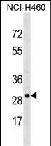
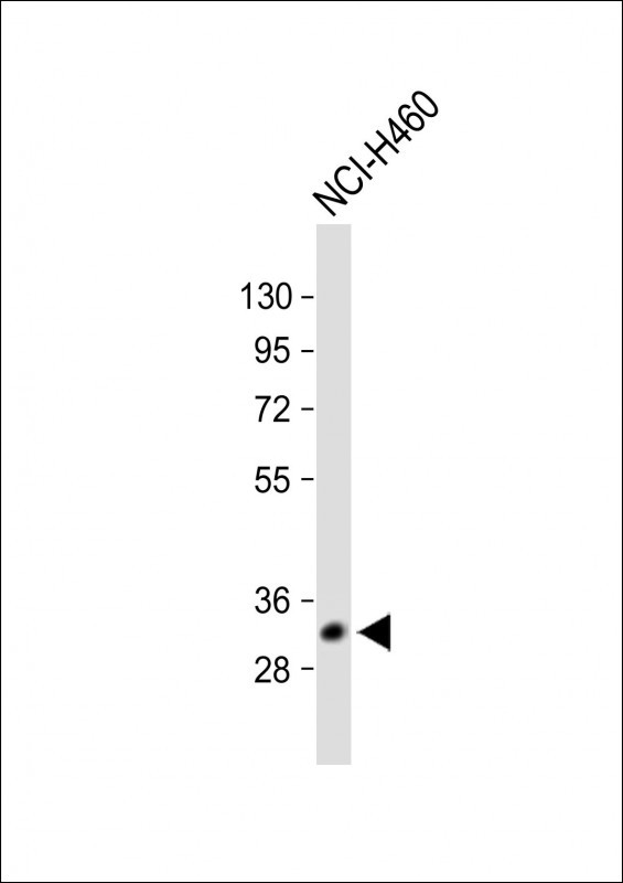
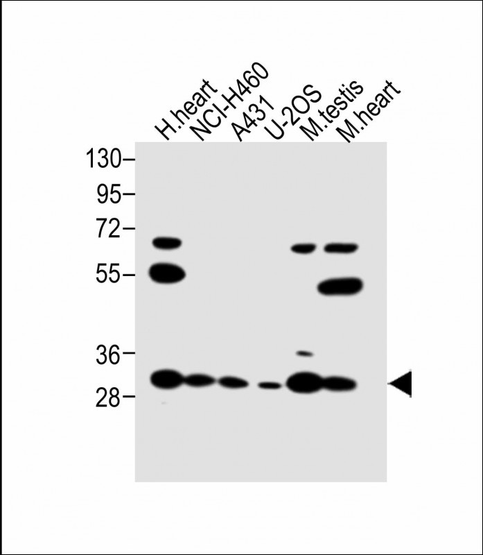
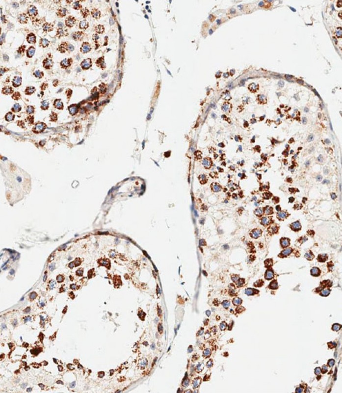
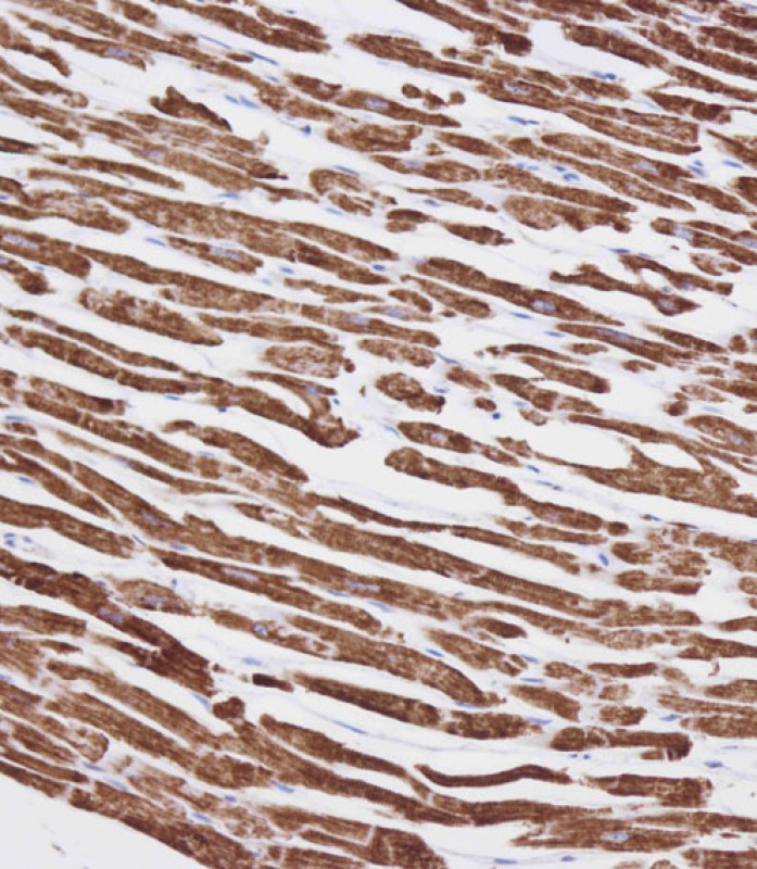
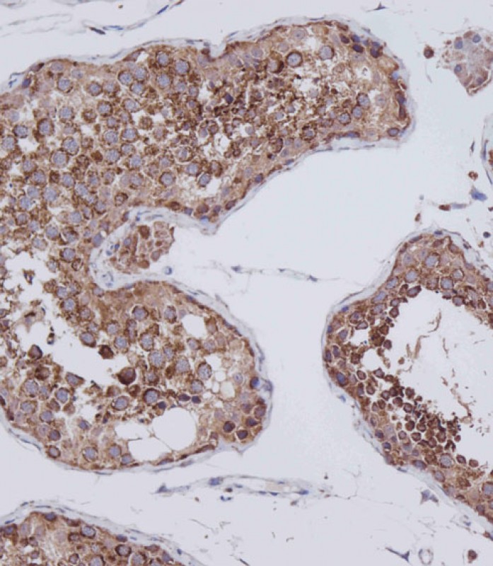
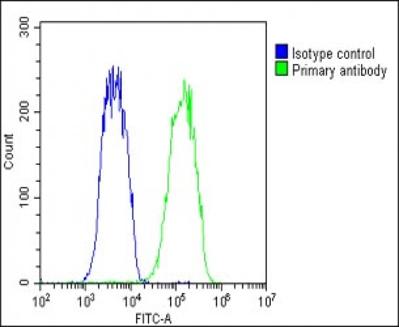
VDAC3 Antibody (Center)
Affinity Purified Rabbit Polyclonal Antibody (Pab)
- SPECIFICATION
- CITATIONS: 1
- PROTOCOLS
- BACKGROUND

Application
| IHC-P, WB, FC, IHC-P-Leica, E |
|---|---|
| Primary Accession | Q9Y277 |
| Other Accession | Q9TT13, Q29380, Q60931, Q9MZ13, NP_005653.3, NP_001129166.1 |
| Reactivity | Human, Mouse, Rat |
| Predicted | Bovine, Pig, Rabbit |
| Host | Rabbit |
| Clonality | Polyclonal |
| Isotype | Rabbit IgG |
| Calculated MW | 30659 Da |
| Antigen Region | 156-183 aa |
| Gene ID | 7419 |
|---|---|
| Other Names | Voltage-dependent anion-selective channel protein 3, VDAC-3, hVDAC3, Outer mitochondrial membrane protein porin 3, VDAC3 |
| Target/Specificity | This VDAC3 antibody is generated from rabbits immunized with a KLH conjugated synthetic peptide between 156-183 amino acids from the Central region of human VDAC3. |
| Dilution | IHC-P~~1:100 WB~~1:1000 FC~~1:25 IHC-P-Leica~~1:500 E~~Use at an assay dependent concentration. |
| Format | Purified polyclonal antibody supplied in PBS with 0.09% (W/V) sodium azide. This antibody is purified through a protein A column, followed by peptide affinity purification. |
| Storage | Maintain refrigerated at 2-8°C for up to 2 weeks. For long term storage store at -20°C in small aliquots to prevent freeze-thaw cycles. |
| Precautions | VDAC3 Antibody (Center) is for research use only and not for use in diagnostic or therapeutic procedures. |
| Name | VDAC3 (HGNC:12674) |
|---|---|
| Function | Non-selective voltage-gated ion channel that mediates the transport of anions and cations through the mitochondrion outer membrane and plasma membrane (PubMed:31935282). Forms a high-conducting channel with a stable open state and a voltage-induced closure with a mild preference for anions over cations (PubMed:31935282). Involved in male fertility and sperm mitochondrial sheath formation (By similarity). |
| Cellular Location | Mitochondrion outer membrane {ECO:0000250|UniProtKB:P21796}. Membrane Note=May localize to non-mitochondrial membranes |
| Tissue Location | Expressed in erythrocytes (at protein level) (PubMed:27641616). Widely expressed. Highest in testis (PubMed:9781040). |

Provided below are standard protocols that you may find useful for product applications.
Background
VDAC3 belongs to a group of mitochondrial membrane channels involved in translocation of adenine nucleotides through the outer membrane. These channels may also function as a mitochondrial binding site for hexokinase (see HK1; MIM 142600) and glycerol kinase (GK; MIM 300474) (Rahmani et al., 1998).[supplied by OMIM].
References
Reina, S., et al. FEBS Lett. 584(13):2837-2844(2010)
Lefievre, L., et al. Proteomics 7(17):3066-3084(2007)
Lamesch, P., et al. Genomics 89(3):307-315(2007)
Ewing, R.M., et al. Mol. Syst. Biol. 3, 89 (2007) :
Rush, J., et al. Nat. Biotechnol. 23(1):94-101(2005)
If you have used an Abcepta product and would like to share how it has performed, please click on the "Submit Review" button and provide the requested information. Our staff will examine and post your review and contact you if needed.
If you have any additional inquiries please email technical services at tech@abcepta.com.
Ordering Information
Other Products
Shipping Information
