


 Foundational characteristics of cancer include proliferation, angiogenesis, migration, evasion of apoptosis, and cellular immortality. Find key markers for these cellular processes and antibodies to detect them.
 The SUMOplot™ Analysis Program predicts and scores sumoylation sites in your protein. SUMOylation is a post-translational modification involved in various cellular processes, such as nuclear-cytosolic transport, transcriptional regulation, apoptosis, protein stability, response to stress, and progression through the cell cycle.
 The Autophagy Receptor Motif Plotter predicts and scores autophagy receptor binding sites in your protein. Identifying proteins connected to this pathway is critical to understanding the role of autophagy in physiological as well as pathological processes such as development, differentiation, neurodegenerative diseases, stress, infection, and cancer.
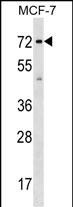
SGOL1 Antibody (Center)
Affinity Purified Rabbit Polyclonal Antibody (Pab)
- SPECIFICATION
- CITATIONS
- PROTOCOLS
- BACKGROUND

Application
| WB, E |
|---|---|
| Primary Accession | Q5FBB7 |
| Other Accession | NP_001012409.1, NP_001012410.1 |
| Reactivity | Human |
| Host | Rabbit |
| Clonality | Polyclonal |
| Isotype | Rabbit IgG |
| Calculated MW | 64190 Da |
| Antigen Region | 339-367 aa |
| Gene ID | 151648 |
|---|---|
| Other Names | Shugoshin-like 1, hSgo1, Serologically defined breast cancer antigen NY-BR-85, SGOL1, SGO1 |
| Target/Specificity | This SGOL1 antibody is generated from rabbits immunized with a KLH conjugated synthetic peptide between 339-367 amino acids from the Central region of human SGOL1. |
| Dilution | WB~~1:1000 E~~Use at an assay dependent concentration. |
| Format | Purified polyclonal antibody supplied in PBS with 0.09% (W/V) sodium azide. This antibody is purified through a protein A column, followed by peptide affinity purification. |
| Storage | Maintain refrigerated at 2-8°C for up to 2 weeks. For long term storage store at -20°C in small aliquots to prevent freeze-thaw cycles. |
| Precautions | SGOL1 Antibody (Center) is for research use only and not for use in diagnostic or therapeutic procedures. |
| Name | SGO1 (HGNC:25088) |
|---|---|
| Synonyms | SGOL1 |
| Function | Plays a central role in chromosome cohesion during mitosis by preventing premature dissociation of cohesin complex from centromeres after prophase, when most of cohesin complex dissociates from chromosomes arms. May act by preventing phosphorylation of the STAG2 subunit of cohesin complex at the centromere, ensuring cohesin persistence at centromere until cohesin cleavage by ESPL1/separase at anaphase. Essential for proper chromosome segregation during mitosis and this function requires interaction with PPP2R1A. Its phosphorylated form is necessary for chromosome congression and for the proper attachment of spindle microtubule to the kinetochore. Necessary for kinetochore localization of PLK1 and CENPF. May play a role in the tension sensing mechanism of the spindle-assembly checkpoint by regulating PLK1 kinetochore affinity. Isoform 3 plays a role in maintaining centriole cohesion involved in controlling spindle pole integrity. Involved in centromeric enrichment of AUKRB in prometaphase. |
| Cellular Location | Nucleus. Chromosome, centromere. Chromosome, centromere, kinetochore. Cytoplasm, cytoskeleton, spindle pole. Cytoplasm, cytoskeleton, microtubule organizing center, centrosome. Nucleus speckle. Note=Localizes to the inner centromere throughout prophase until metaphase and disappears at anaphase (PubMed:16541025). Centromeric localization requires the presence of BUB1 and the interaction with PPP2R1A (PubMed:15604152, PubMed:16541025, PubMed:16580887). Colocalizes with NEK2 at the kinetochore (PubMed:17621308). Colocalizes with and SS18L1 at the kinetochore (PubMed:16582621). Phosphorylation by AUKRB and the presence of BUB1 are required for localization to the kinetochore (PubMed:17617734). Isoform 1 primarily localizes to kinetochores during G2 phase and mitotic prophase, metaphase, and anaphase and does not appear to be associated with kinetochores during late mitosis (PubMed:16582621). Isoform 3 is found at the centrosome in interphase and at spindle poles in mitosis and its spindle pole localization is PLK1 dependent (PubMed:16582621). Isoform 3 does not localize to kinetochores during any stages of the cell cycle (PubMed:16582621) |
| Tissue Location | Widely expressed. Highly expressed in testis. Expressed in lung, small intestine, breast, liver and placenta Strongly overexpressed in 90% of breast cancers tested |

Thousands of laboratories across the world have published research that depended on the performance of antibodies from Abcepta to advance their research. Check out links to articles that cite our products in major peer-reviewed journals, organized by research category.
info@abcepta.com, and receive a free "I Love Antibodies" mug.

Provided below are standard protocols that you may find useful for product applications.
Background
SGOL1 plays a central role in chromosome cohesion during mitosis by preventing premature dissociation of cohesin complex from centromeres after prophase, when most of cohesin complex dissociates from chromosomes arms. May act by preventing phosphorylation of the STAG2 subunit of cohesin complex at the centromere, ensuring cohesin persistence at centromere until cohesin cleavage by ESPL1/separase at anaphase. Essential for proper chromosome segregation during mitosis and this function requires interaction with PPP2R1A. Its phosphorylated form is necessary for chromosome congression and for the proper attachment of spindle microtubule to the kinetochore. Necessary for kinetochore localization of PLK1 and CENPF. May play a role in the tension sensing mechanism of the spindle-assembly checkpoint by regulating PLK1 kinetochore affinity. Isoform 3 plays a role in maintaining centriole cohesion involved in controlling spindle pole integrity.
References
Rose, J.E., et al. Mol. Med. 16 (7-8), 247-253 (2010) :
Okamoto, N., et al. Genes Cells 15(5):471-484(2010)
Xu, Z., et al. Mol. Cell 35(4):426-441(2009)
Gambe, A.E., et al. FEBS Lett. 583(12):1951-1956(2009)
Karamysheva, Z., et al. J. Biol. Chem. 284(3):1772-1780(2009)
If you have used an Abcepta product and would like to share how it has performed, please click on the "Submit Review" button and provide the requested information. Our staff will examine and post your review and contact you if needed.
If you have any additional inquiries please email technical services at tech@abcepta.com.
Ordering Information
Shipping Information
