


 Foundational characteristics of cancer include proliferation, angiogenesis, migration, evasion of apoptosis, and cellular immortality. Find key markers for these cellular processes and antibodies to detect them.
 The SUMOplot™ Analysis Program predicts and scores sumoylation sites in your protein. SUMOylation is a post-translational modification involved in various cellular processes, such as nuclear-cytosolic transport, transcriptional regulation, apoptosis, protein stability, response to stress, and progression through the cell cycle.
 The Autophagy Receptor Motif Plotter predicts and scores autophagy receptor binding sites in your protein. Identifying proteins connected to this pathway is critical to understanding the role of autophagy in physiological as well as pathological processes such as development, differentiation, neurodegenerative diseases, stress, infection, and cancer.
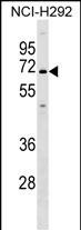
KCNN1 Antibody (C-term)
Affinity Purified Rabbit Polyclonal Antibody (Pab)
- SPECIFICATION
- CITATIONS
- PROTOCOLS
- BACKGROUND

Application
| WB, E |
|---|---|
| Primary Accession | Q92952 |
| Other Accession | NP_002239.2 |
| Reactivity | Human |
| Host | Rabbit |
| Clonality | Polyclonal |
| Isotype | Rabbit IgG |
| Calculated MW | 59987 Da |
| Antigen Region | 391-419 aa |
| Gene ID | 3780 |
|---|---|
| Other Names | Small conductance calcium-activated potassium channel protein 1, SK1, SKCa 1, SKCa1, KCa21, KCNN1, SK |
| Target/Specificity | This KCNN1 antibody is generated from rabbits immunized with a KLH conjugated synthetic peptide between 391-419 amino acids from the C-terminal region of human KCNN1. |
| Dilution | WB~~1:1000 E~~Use at an assay dependent concentration. |
| Format | Purified polyclonal antibody supplied in PBS with 0.09% (W/V) sodium azide. This antibody is purified through a protein A column, followed by peptide affinity purification. |
| Storage | Maintain refrigerated at 2-8°C for up to 2 weeks. For long term storage store at -20°C in small aliquots to prevent freeze-thaw cycles. |
| Precautions | KCNN1 Antibody (C-term) is for research use only and not for use in diagnostic or therapeutic procedures. |
| Name | KCNN1 {ECO:0000303|PubMed:10516439, ECO:0000312|HGNC:HGNC:6290} |
|---|---|
| Function | Small conductance calcium-activated potassium channel that mediates the voltage-independent transmembrane transfer of potassium across the cell membrane through a constitutive interaction with calmodulin which binds the intracellular calcium allowing its opening (PubMed:17142458, PubMed:8781233, PubMed:9287325). The current is characterized by a voltage-independent activation, an intracellular calcium concentration increase-dependent activation and a single- channel conductance of about 3 picosiemens (PubMed:8781233). Also presents an inwardly rectifying current, thus reducing its already small outward conductance of potassium ions, which is particularly the case when the membrane potential displays positive values, above + 20 mV (Probable). Activation is followed by membrane hyperpolarization (By similarity). Thought to regulate neuronal excitability by contributing to the slow component of synaptic afterhyperpolarization (By similarity). |
| Cellular Location | Membrane; Multi-pass membrane protein. Cytoplasm, myofibril, sarcomere, Z line {ECO:0000250|UniProtKB:Q9EQR3} |

Thousands of laboratories across the world have published research that depended on the performance of antibodies from Abcepta to advance their research. Check out links to articles that cite our products in major peer-reviewed journals, organized by research category.
info@abcepta.com, and receive a free "I Love Antibodies" mug.

Provided below are standard protocols that you may find useful for product applications.
Background
Action potentials in vertebrate neurons are followed by an afterhyperpolarization (AHP) that may persist for several seconds and may have profound consequences for the firing pattern of the neuron. Each component of the AHP is kinetically distinct and is mediated by different calcium-activated potassium channels. The protein encoded by this gene is activated before membrane hyperpolarization and is thought to regulate neuronal excitability by contributing to the slow component of synaptic AHP. The encoded protein is an integral membrane protein that forms a voltage-independent calcium-activated channel with three other calmodulin-binding subunits. This gene is a member of the KCNN family of potassium channel genes.
References
Wu, C., et al. Proteomics 7(11):1775-1785(2007)
Wei, A.D., et al. Pharmacol. Rev. 57(4):463-472(2005)
Arnold, S.J., et al. Neuroreport 14(2):191-195(2003)
Boettger, M.K., et al. Brain 125 (PT 2), 252-263 (2002) :
Zhang, B.M., et al. Biochemistry 40(10):3189-3195(2001)
If you have used an Abcepta product and would like to share how it has performed, please click on the "Submit Review" button and provide the requested information. Our staff will examine and post your review and contact you if needed.
If you have any additional inquiries please email technical services at tech@abcepta.com.
Ordering Information
Shipping Information
