


 Foundational characteristics of cancer include proliferation, angiogenesis, migration, evasion of apoptosis, and cellular immortality. Find key markers for these cellular processes and antibodies to detect them.
 The SUMOplot™ Analysis Program predicts and scores sumoylation sites in your protein. SUMOylation is a post-translational modification involved in various cellular processes, such as nuclear-cytosolic transport, transcriptional regulation, apoptosis, protein stability, response to stress, and progression through the cell cycle.
 The Autophagy Receptor Motif Plotter predicts and scores autophagy receptor binding sites in your protein. Identifying proteins connected to this pathway is critical to understanding the role of autophagy in physiological as well as pathological processes such as development, differentiation, neurodegenerative diseases, stress, infection, and cancer.
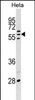
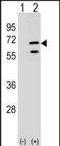
POLR3C Antibody (Center)
Affinity Purified Rabbit Polyclonal Antibody (Pab)
- SPECIFICATION
- CITATIONS
- PROTOCOLS
- BACKGROUND

Application
| WB, E |
|---|---|
| Primary Accession | Q9BUI4 |
| Other Accession | NP_006459.3 |
| Reactivity | Human |
| Host | Rabbit |
| Clonality | Polyclonal |
| Isotype | Rabbit IgG |
| Calculated MW | 60612 Da |
| Antigen Region | 202-230 aa |
| Gene ID | 10623 |
|---|---|
| Other Names | DNA-directed RNA polymerase III subunit RPC3, RNA polymerase III subunit C3, DNA-directed RNA polymerase III subunit C, RNA polymerase III 62 kDa subunit, RPC62, POLR3C |
| Target/Specificity | This POLR3C antibody is generated from rabbits immunized with a KLH conjugated synthetic peptide between 202-230 amino acids from the Central region of human POLR3C. |
| Dilution | WB~~1:1000 E~~Use at an assay dependent concentration. |
| Format | Purified polyclonal antibody supplied in PBS with 0.09% (W/V) sodium azide. This antibody is purified through a protein A column, followed by peptide affinity purification. |
| Storage | Maintain refrigerated at 2-8°C for up to 2 weeks. For long term storage store at -20°C in small aliquots to prevent freeze-thaw cycles. |
| Precautions | POLR3C Antibody (Center) is for research use only and not for use in diagnostic or therapeutic procedures. |
| Name | POLR3C (HGNC:30076) |
|---|---|
| Function | DNA-dependent RNA polymerase catalyzes the transcription of DNA into RNA using the four ribonucleoside triphosphates as substrates (PubMed:20413673, PubMed:33558764, PubMed:33558766, PubMed:34675218, PubMed:35637192). Specific peripheric component of RNA polymerase III (Pol III) which synthesizes small non-coding RNAs including 5S rRNA, snRNAs, tRNAs and miRNAs from at least 500 distinct genomic loci (PubMed:20413673, PubMed:33558764, PubMed:33558766, PubMed:35637192). Part of POLR3C/RPC3-POLR3F/RPC6-POLR3G/RPC7 heterotrimer, coordinates the dynamics of Pol III stalk and clamp modules during the transition from apo to elongation state (PubMed:33558764, PubMed:33558766). Pol III plays a key role in sensing and limiting infection by intracellular bacteria and DNA viruses. Acts as a nuclear and cytosolic DNA sensor involved in innate immune response. Can sense non-self dsDNA that serves as template for transcription into dsRNA. The non-self RNA polymerase III transcripts, such as Epstein-Barr virus-encoded RNAs (EBERs) induce type I interferon and NF-kappa-B through the RIG-I pathway (PubMed:19609254, PubMed:19631370). Preferentially binds single-stranded DNA (ssDNA) in a sequence-independent manner (PubMed:21358628). |
| Cellular Location | Nucleus. |

Thousands of laboratories across the world have published research that depended on the performance of antibodies from Abcepta to advance their research. Check out links to articles that cite our products in major peer-reviewed journals, organized by research category.
info@abcepta.com, and receive a free "I Love Antibodies" mug.

Provided below are standard protocols that you may find useful for product applications.
Background
DNA-dependent RNA polymerase catalyzes the transcription of DNA into RNA using the four ribonucleoside triphosphates as substrates. Specific core component of RNA polymerase III which synthesizes small RNAs, such as 5S rRNA and tRNAs. May direct with other members of the subcomplex RNA Pol III binding to the TFIIIB-DNA complex via the interactions between TFIIIB and POLR3F. May be involved either in the recruitment and stabilization of the subcomplex within RNA polymerase III, or in stimulating catalytic functions of other subunits during initiation. Plays a key role in sensing and limiting infection by intracellular bacteria and DNA viruses. Acts as nuclear and cytosolic DNA sensor involved in innate immune response. Can sense non-self dsDNA that serves as template for transcription into dsRNA. The non-self RNA polymerase III transcripts, such as Epstein-Barr virus-encoded RNAs (EBERs) induce type I interferon and NF-Kappa-B through the RIG-I pathway.
References
Ablasser, A., et al. Nat. Immunol. 10(10):1065-1072(2009)
Hu, P., et al. Mol. Cell. Biol. 22(22):8044-8055(2002)
Kuwana, M., et al. Arthritis Rheum. 46(10):2742-2747(2002)
Hsieh, Y.J., et al. Mol. Cell. Biol. 19(11):7697-7704(1999)
Hsieh, Y.J., et al. Mol. Cell. Biol. 19(7):4944-4952(1999)
If you have used an Abcepta product and would like to share how it has performed, please click on the "Submit Review" button and provide the requested information. Our staff will examine and post your review and contact you if needed.
If you have any additional inquiries please email technical services at tech@abcepta.com.
Ordering Information
Other Products
Shipping Information
