


 Foundational characteristics of cancer include proliferation, angiogenesis, migration, evasion of apoptosis, and cellular immortality. Find key markers for these cellular processes and antibodies to detect them.
 The SUMOplot™ Analysis Program predicts and scores sumoylation sites in your protein. SUMOylation is a post-translational modification involved in various cellular processes, such as nuclear-cytosolic transport, transcriptional regulation, apoptosis, protein stability, response to stress, and progression through the cell cycle.
 The Autophagy Receptor Motif Plotter predicts and scores autophagy receptor binding sites in your protein. Identifying proteins connected to this pathway is critical to understanding the role of autophagy in physiological as well as pathological processes such as development, differentiation, neurodegenerative diseases, stress, infection, and cancer.
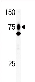
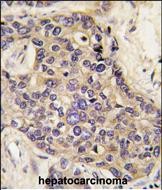
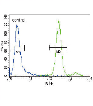
HGF Antibody (C-term)
Purified Rabbit Polyclonal Antibody (Pab)
- SPECIFICATION
- CITATIONS: 2
- PROTOCOLS
- BACKGROUND

Application
| IHC-P, WB, FC, E |
|---|---|
| Primary Accession | P14210 |
| Reactivity | Human |
| Host | Rabbit |
| Clonality | Polyclonal |
| Isotype | Rabbit IgG |
| Calculated MW | 83134 Da |
| Antigen Region | 521-554 aa |
| Gene ID | 3082 |
|---|---|
| Other Names | Hepatocyte growth factor, Hepatopoietin-A, Scatter factor, SF, Hepatocyte growth factor alpha chain, Hepatocyte growth factor beta chain, HGF, HPTA |
| Target/Specificity | This HGF antibody is generated from rabbits immunized with a KLH conjugated synthetic peptide between 521-554 amino acids from the C-terminal region of human HGF. |
| Dilution | IHC-P~~1:50~100 WB~~1:1000 FC~~1:10~50 E~~Use at an assay dependent concentration. |
| Format | Purified polyclonal antibody supplied in PBS with 0.09% (W/V) sodium azide. This antibody is prepared by Saturated Ammonium Sulfate (SAS) precipitation followed by dialysis against PBS. |
| Storage | Maintain refrigerated at 2-8°C for up to 2 weeks. For long term storage store at -20°C in small aliquots to prevent freeze-thaw cycles. |
| Precautions | HGF Antibody (C-term) is for research use only and not for use in diagnostic or therapeutic procedures. |
| Name | HGF |
|---|---|
| Synonyms | HPTA |
| Function | Potent mitogen for mature parenchymal hepatocyte cells, seems to be a hepatotrophic factor, and acts as a growth factor for a broad spectrum of tissues and cell types (PubMed:20624990). Activating ligand for the receptor tyrosine kinase MET by binding to it and promoting its dimerization (PubMed:15167892, PubMed:20977675). Activates MAPK signaling following TMPRSS13 cleavage and activation (PubMed:20977675). |

Provided below are standard protocols that you may find useful for product applications.
Background
Hepatocyte growth factor regulates cell growth, cell motility, and morphogenesis by activating a tyrosine kinase signaling cascade after binding to the proto-oncogenic c-Met receptor. Hepatocyte growth factor is secreted by mesenchymal cells and acts as a multi-functional cytokine on cells of mainly epithelial origin. Its ability to stimulate mitogenesis, cell motility, and matrix invasion gives it a central role in angiogenesis, tumorogenesis, and tissue regeneration. It is secreted as a single inactive polypeptide and is cleaved by serine proteases into a 69-kDa alpha-chain and 34-kDa beta-chain. A disulfide bond between the alpha and beta chains produces the active, heterodimeric molecule. The protein belongs to the plasminogen subfamily of S1 peptidases but has no detectable protease activity. Alternative splicing of this gene produces multiple transcript variants encoding different isoforms. Transcript Variant: This variant (1) encodes the longest isoform (1). To date, experimental evidence for cleavage of the proprotein into two mature chains has been shown only for isoform 1.
References
Ryugo, M., et al., Transplantation 78(8):1153-1158 (2004).
Lyon, M., et al., J. Biol. Chem. 279(42):43560-43567 (2004).
He, Y., et al., World J. Gastroenterol. 10(19):2827-2830 (2004).
Tjin, E.P., et al., Blood 104(7):2172-2175 (2004).
Matsuda-Hashii, Y., et al., Exp. Hematol. 32(10):955-961 (2004).
If you have used an Abcepta product and would like to share how it has performed, please click on the "Submit Review" button and provide the requested information. Our staff will examine and post your review and contact you if needed.
If you have any additional inquiries please email technical services at tech@abcepta.com.
Ordering Information
Other Products
Shipping Information
