


 Foundational characteristics of cancer include proliferation, angiogenesis, migration, evasion of apoptosis, and cellular immortality. Find key markers for these cellular processes and antibodies to detect them.
 The SUMOplot™ Analysis Program predicts and scores sumoylation sites in your protein. SUMOylation is a post-translational modification involved in various cellular processes, such as nuclear-cytosolic transport, transcriptional regulation, apoptosis, protein stability, response to stress, and progression through the cell cycle.
 The Autophagy Receptor Motif Plotter predicts and scores autophagy receptor binding sites in your protein. Identifying proteins connected to this pathway is critical to understanding the role of autophagy in physiological as well as pathological processes such as development, differentiation, neurodegenerative diseases, stress, infection, and cancer.
PIGO Antibody (C-term)
Affinity Purified Rabbit Polyclonal Antibody (Pab)
- SPECIFICATION
- CITATIONS
- PROTOCOLS
- BACKGROUND

Application
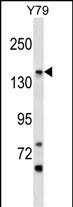
| WB, E |
|---|---|
| Primary Accession | Q8TEQ8 |
| Other Accession | NP_116023.2, NP_690577.2 |
| Reactivity | Human |
| Host | Rabbit |
| Clonality | Polyclonal |
| Isotype | Rabbit IgG |
| Calculated MW | 118699 Da |
| Antigen Region | 955-983 aa |
| Gene ID | 84720 |
|---|---|
| Other Names | GPI ethanolamine phosphate transferase 3, 2---, Phosphatidylinositol-glycan biosynthesis class O protein, PIG-O, PIGO |
| Target/Specificity | This PIGO antibody is generated from rabbits immunized with a KLH conjugated synthetic peptide between 955-983 amino acids from the C-terminal region of human PIGO. |
| Dilution | WB~~1:1000 E~~Use at an assay dependent concentration. |
| Format | Purified polyclonal antibody supplied in PBS with 0.09% (W/V) sodium azide. This antibody is purified through a protein A column, followed by peptide affinity purification. |
| Storage | Maintain refrigerated at 2-8°C for up to 2 weeks. For long term storage store at -20°C in small aliquots to prevent freeze-thaw cycles. |
| Precautions | PIGO Antibody (C-term) is for research use only and not for use in diagnostic or therapeutic procedures. |
| Name | PIGO (HGNC:23215) |
|---|---|
| Function | Catalytic subunit of the ethanolamine phosphate transferase 3 complex that transfers an ethanolamine phosphate (EtNP) from a phosphatidylethanolamine (PE) to the 6-OH position of the third alpha- 1,2-linked mannose of the a 2-acyl-6-[alpha-D-mannosyl-(1->2)-alpha-D- mannosyl-(1->6)-2-phosphoethanolamine-alpha-D-mannosyl-(1->4)-alpha-D- glucosaminyl]-1-(1-radyl,2-acyl-sn-glycero-3-phospho)-1D-myo-inositol (also termed H6) intermediate to generate a a 2-acyl-6-[6- phosphoethanolamine-alpha-D-mannosyl-(1->2)-alpha-D-mannosyl-(1->6)-2- phosphoethanolamine-alpha-D-mannosyl-(1->4)-alpha-D-glucosaminyl]-1-(1- radyl,2-acyl-sn-glycero-3-phospho)-1D-myo-inositol (also termed H7) and participates in the tenth step of the glycosylphosphatidylinositol- anchor biosynthesis. |
| Cellular Location | Endoplasmic reticulum membrane {ECO:0000250|UniProtKB:Q9JJI6}; Multi-pass membrane protein |

Thousands of laboratories across the world have published research that depended on the performance of antibodies from Abcepta to advance their research. Check out links to articles that cite our products in major peer-reviewed journals, organized by research category.
info@abcepta.com, and receive a free "I Love Antibodies" mug.

Provided below are standard protocols that you may find useful for product applications.
Background
This gene encodes a protein that is involved in glycosylphosphatidylinositol (GPI)-anchor biosynthesis. The GPI-anchor is a glycolipid which contains three mannose molecules in its core backbone. The GPI-anchor is found on many blood cells and serves to anchor proteins to the cell surface. This protein is involved in the transfer of ethanolaminephosphate (EtNP) to the third mannose in GPI. At least two alternatively spliced transcripts encoding distinct isoforms have been found for this gene.
References
Bailey, S.D., et al. Diabetes Care (2010) In press :
Rose, J.E., et al. Mol. Med. 16 (7-8), 247-253 (2010) :
Talmud, P.J., et al. Am. J. Hum. Genet. 85(5):628-642(2009)
Humphray, S.J., et al. Nature 429(6990):369-374(2004)
Clark, H.F., et al. Genome Res. 13(10):2265-2270(2003)
If you have used an Abcepta product and would like to share how it has performed, please click on the "Submit Review" button and provide the requested information. Our staff will examine and post your review and contact you if needed.
If you have any additional inquiries please email technical services at tech@abcepta.com.
Ordering Information
Shipping Information
