


 Foundational characteristics of cancer include proliferation, angiogenesis, migration, evasion of apoptosis, and cellular immortality. Find key markers for these cellular processes and antibodies to detect them.
 The SUMOplot™ Analysis Program predicts and scores sumoylation sites in your protein. SUMOylation is a post-translational modification involved in various cellular processes, such as nuclear-cytosolic transport, transcriptional regulation, apoptosis, protein stability, response to stress, and progression through the cell cycle.
 The Autophagy Receptor Motif Plotter predicts and scores autophagy receptor binding sites in your protein. Identifying proteins connected to this pathway is critical to understanding the role of autophagy in physiological as well as pathological processes such as development, differentiation, neurodegenerative diseases, stress, infection, and cancer.
AP4B1 Antibody (Center)
Affinity Purified Rabbit Polyclonal Antibody (Pab)
- SPECIFICATION
- CITATIONS
- PROTOCOLS
- BACKGROUND

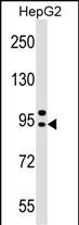
Application
| WB, E |
|---|---|
| Primary Accession | Q9Y6B7 |
| Other Accession | Q9WV76, NP_006585.2 |
| Reactivity | Human |
| Predicted | Mouse |
| Host | Rabbit |
| Clonality | Polyclonal |
| Isotype | Rabbit IgG |
| Calculated MW | 83260 Da |
| Antigen Region | 492-520 aa |
| Gene ID | 10717 |
|---|---|
| Other Names | AP-4 complex subunit beta-1, AP-4 adaptor complex subunit beta, Adaptor-related protein complex 4 subunit beta-1, Beta subunit of AP-4, Beta4-adaptin, AP4B1 |
| Target/Specificity | This AP4B1 antibody is generated from rabbits immunized with a KLH conjugated synthetic peptide between 492-520 amino acids from the Central region of human AP4B1. |
| Dilution | WB~~1:1000 E~~Use at an assay dependent concentration. |
| Format | Purified polyclonal antibody supplied in PBS with 0.09% (W/V) sodium azide. This antibody is purified through a protein A column, followed by peptide affinity purification. |
| Storage | Maintain refrigerated at 2-8°C for up to 2 weeks. For long term storage store at -20°C in small aliquots to prevent freeze-thaw cycles. |
| Precautions | AP4B1 Antibody (Center) is for research use only and not for use in diagnostic or therapeutic procedures. |
| Name | AP4B1 (HGNC:572) |
|---|---|
| Function | Component of the adaptor protein complex 4 (AP-4). Adaptor protein complexes are vesicle coat components involved both in vesicle formation and cargo selection. They control the vesicular transport of proteins in different trafficking pathways (PubMed:10066790, PubMed:10436028). AP-4 forms a non clathrin-associated coat on vesicles departing the trans-Golgi network (TGN) and may be involved in the targeting of proteins from the trans-Golgi network (TGN) to the endosomal-lysosomal system. It is also involved in protein sorting to the basolateral membrane in epithelial cells and the proper asymmetric localization of somatodendritic proteins in neurons. AP-4 is involved in the recognition and binding of tyrosine-based sorting signals found in the cytoplasmic part of cargos, but may also recognize other types of sorting signal (Probable). |
| Cellular Location | Golgi apparatus, trans-Golgi network membrane; Peripheral membrane protein |
| Tissue Location | Widely expressed.. |

Thousands of laboratories across the world have published research that depended on the performance of antibodies from Abcepta to advance their research. Check out links to articles that cite our products in major peer-reviewed journals, organized by research category.
info@abcepta.com, and receive a free "I Love Antibodies" mug.

Provided below are standard protocols that you may find useful for product applications.
Background
The heterotetrameric adaptor protein (AP) complexes sort integral membrane proteins at various stages of the endocytic and secretory pathways. AP4 is composed of 2 large chains, beta-4 (AP4B1) and epsilon-4 (AP4E1; MIM 607244), a medium chain, mu-4 (AP4M1; MIM 602296), and a small chain, sigma-4 (AP4S1; MIM 607243).
References
Ewing, R.M., et al. Mol. Syst. Biol. 3, 89 (2007) :
Cayrol, C., et al. Biochem. Biophys. Res. Commun. 298(5):720-730(2002)
Takatsu, H., et al. Biochem. Biophys. Res. Commun. 284(4):1083-1089(2001)
Hirst, J., et al. Mol. Biol. Cell 10(8):2787-2802(1999)
Dell'Angelica, E.C., et al. J. Biol. Chem. 274(11):7278-7285(1999)
If you have used an Abcepta product and would like to share how it has performed, please click on the "Submit Review" button and provide the requested information. Our staff will examine and post your review and contact you if needed.
If you have any additional inquiries please email technical services at tech@abcepta.com.
Ordering Information
Other Products
Shipping Information
