


 Foundational characteristics of cancer include proliferation, angiogenesis, migration, evasion of apoptosis, and cellular immortality. Find key markers for these cellular processes and antibodies to detect them.
 The SUMOplot™ Analysis Program predicts and scores sumoylation sites in your protein. SUMOylation is a post-translational modification involved in various cellular processes, such as nuclear-cytosolic transport, transcriptional regulation, apoptosis, protein stability, response to stress, and progression through the cell cycle.
 The Autophagy Receptor Motif Plotter predicts and scores autophagy receptor binding sites in your protein. Identifying proteins connected to this pathway is critical to understanding the role of autophagy in physiological as well as pathological processes such as development, differentiation, neurodegenerative diseases, stress, infection, and cancer.
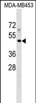
MR1 Antibody (C-term)
Affinity Purified Rabbit Polyclonal Antibody (Pab)
- SPECIFICATION
- CITATIONS
- PROTOCOLS
- BACKGROUND

Application
| WB, E |
|---|---|
| Primary Accession | Q95460 |
| Other Accession | NP_001181929.1, NP_001181928.1 |
| Reactivity | Human |
| Host | Rabbit |
| Clonality | Polyclonal |
| Isotype | Rabbit IgG |
| Calculated MW | 39366 Da |
| Antigen Region | 312-341 aa |
| Gene ID | 3140 |
|---|---|
| Other Names | Major histocompatibility complex class I-related gene protein, MHC class I-related gene protein, Class I histocompatibility antigen-like protein, MR1 |
| Target/Specificity | This MR1 antibody is generated from rabbits immunized with a KLH conjugated synthetic peptide between 312-341 amino acids from the C-terminal region of human MR1. |
| Dilution | WB~~1:1000 E~~Use at an assay dependent concentration. |
| Format | Purified polyclonal antibody supplied in PBS with 0.09% (W/V) sodium azide. This antibody is purified through a protein A column, followed by peptide affinity purification. |
| Storage | Maintain refrigerated at 2-8°C for up to 2 weeks. For long term storage store at -20°C in small aliquots to prevent freeze-thaw cycles. |
| Precautions | MR1 Antibody (C-term) is for research use only and not for use in diagnostic or therapeutic procedures. |
| Name | MR1 {ECO:0000303|PubMed:19416870, ECO:0000312|HGNC:HGNC:4975} |
|---|---|
| Function | Antigen-presenting molecule specialized in displaying microbial pyrimidine-based metabolites to alpha-beta T cell receptors (TCR) on innate-type mucosal-associated invariant T (MAIT) cells (PubMed:19416870, PubMed:23457030, PubMed:22692454, PubMed:23051753, PubMed:24101382, PubMed:23846752, PubMed:26795251). In complex with B2M preferentially presents riboflavin-derived metabolites to semi- invariant TRAV1.2 TCRs on MAIT cells, guiding immune surveillance of the microbial metabolome at mucosal epithelial barriers (PubMed:20581831, PubMed:24101382, PubMed:24695216, PubMed:26795251). Signature pyrimidine-based microbial antigens are generated via non- enzymatic condensation of metabolite intermediates of the riboflavin pathway with by-products arising from other metabolic pathways such as glycolysis. Typical potent antigenic metabolites are 5-(2- oxoethylideneamino)-6-D-ribitylaminouracil (5-OE-RU) and 5-(2- oxopropylideneamino)-6-D-ribitylaminouracil (5-OP-RU), products of condensation of 5-amino-6-D-ribityaminouracil (5-A-RU) with glyoxal or methylglyoxal by-products, respectively (PubMed:24695216, PubMed:32958637, PubMed:32709702). May present microbial antigens to various TRAV1-2-negative MAIT cell subsets, providing for unique recognition of diverse microbes, including pathogens that do not synthesize riboflavin (PubMed:27527800, PubMed:31113973). Upon antigen recognition, elicits rapid innate-type MAIT cell activation to eliminate pathogenic microbes by directly killing infected cells (PubMed:23846752, PubMed:24695216, PubMed:27527800). During T cell development, drives thymic selection and post-thymic terminal differentiation of MAIT cells in a process dependent on commensal microflora (By similarity). Acts as an immune sensor of cancer cell metabolome (PubMed:31959982). May present a tumor-specific or -associated metabolite essential for cancer cell survival to a 'pan- cancer' TCR consisting of TRAV38.2-DV8*TRAJ31 alpha chain paired with a TRBV25.1*TRBJ2.3 beta chain on a non-MAIT CD8-positive T cell clone (MC.7.G5), triggering T cell-mediated killing of a wide range of cancer cell types (PubMed:31959982). |
| Cellular Location | Cell membrane; Single-pass type I membrane protein Endoplasmic reticulum membrane; Single-pass type I membrane protein. Golgi apparatus membrane; Single-pass type I membrane protein. Early endosome membrane; Single-pass type I membrane protein. Late endosome membrane; Single-pass type I membrane protein. Note=In the absence of antigen remains within the endoplasmic reticulum where it acts as a metabolite sensor. Antigen binding triggers trafficking of the ternary complex to the plasma membrane. After presentation, most of these complexes are rapidly internalized and degraded via endocytosis. A small subset recycles via endosomes back to the plasma membrane and may thus acquire and present new antigens that do not efficiently reach the endoplasmic reticulum. [Isoform 3]: Cell membrane; Single-pass type I membrane protein. Endoplasmic reticulum membrane; Single-pass membrane protein. Note=The larger proportion remains in the ER in an immature state. The subset that reach cell surface does it through a B2M-independent pathway. |
| Tissue Location | Ubiquitous (PubMed:7624800, PubMed:9780177). Low expression is detected in peripheral blood B cells, T cells, monocytes and in bronchial epithelial cells (at protein level) (PubMed:27043408) Expressed in plasmablasts or plasma B cells in the lamina propria of ileum, appendix and colon (at protein level) (PubMed:19760593). Highly expressed on a subset of CD45-positive CD3-positive thymocytes (at protein level) (PubMed:22692454). |

Thousands of laboratories across the world have published research that depended on the performance of antibodies from Abcepta to advance their research. Check out links to articles that cite our products in major peer-reviewed journals, organized by research category.
info@abcepta.com, and receive a free "I Love Antibodies" mug.

Provided below are standard protocols that you may find useful for product applications.
Background
MR1 has antigen presentation function. Involved in the development and expansion of a small population of T cells expressing an invariant T cell receptor alpha chain called mucosal-associated invariant T cells (MAIT). MAIT cells are preferentially located in the gut lamina propria and therfore may be involed in monitoring commensal flora or serve as a distress signal. Expression and MAIT cell recognition seem to be ligand-dependent.
References
Gozalbo-Lopez, B., et al. Histol. Histopathol. 24(11):1439-1449(2009)
Stumpf, A.N., et al. Blood 114(17):3684-3692(2009)
Huang, S., et al. Proc. Natl. Acad. Sci. U.S.A. 106(20):8290-8295(2009)
Aldemir, H. Biochem. Biophys. Res. Commun. 366(2):328-334(2008)
Miley, M.J., et al. J. Immunol. 170(12):6090-6098(2003)
If you have used an Abcepta product and would like to share how it has performed, please click on the "Submit Review" button and provide the requested information. Our staff will examine and post your review and contact you if needed.
If you have any additional inquiries please email technical services at tech@abcepta.com.
Ordering Information
Shipping Information
