


 Foundational characteristics of cancer include proliferation, angiogenesis, migration, evasion of apoptosis, and cellular immortality. Find key markers for these cellular processes and antibodies to detect them.
 The SUMOplot™ Analysis Program predicts and scores sumoylation sites in your protein. SUMOylation is a post-translational modification involved in various cellular processes, such as nuclear-cytosolic transport, transcriptional regulation, apoptosis, protein stability, response to stress, and progression through the cell cycle.
 The Autophagy Receptor Motif Plotter predicts and scores autophagy receptor binding sites in your protein. Identifying proteins connected to this pathway is critical to understanding the role of autophagy in physiological as well as pathological processes such as development, differentiation, neurodegenerative diseases, stress, infection, and cancer.
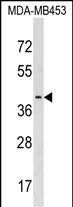
PAFAH2 Antibody (Center)
Affinity Purified Rabbit Polyclonal Antibody (Pab)
- SPECIFICATION
- CITATIONS
- PROTOCOLS
- BACKGROUND

Application
| WB, E |
|---|---|
| Primary Accession | Q99487 |
| Other Accession | Q8VDG7, NP_000428.2 |
| Reactivity | Human |
| Predicted | Mouse |
| Host | Rabbit |
| Clonality | Polyclonal |
| Isotype | Rabbit IgG |
| Calculated MW | 44036 Da |
| Antigen Region | 249-276 aa |
| Gene ID | 5051 |
|---|---|
| Other Names | Platelet-activating factor acetylhydrolase 2, cytoplasmic, Serine-dependent phospholipase A2, SD-PLA2, hSD-PLA2, PAFAH2 |
| Target/Specificity | This PAFAH2 antibody is generated from rabbits immunized with a KLH conjugated synthetic peptide between 249-276 amino acids from the Central region of human PAFAH2. |
| Dilution | WB~~1:1000 E~~Use at an assay dependent concentration. |
| Format | Purified polyclonal antibody supplied in PBS with 0.09% (W/V) sodium azide. This antibody is purified through a protein A column, followed by peptide affinity purification. |
| Storage | Maintain refrigerated at 2-8°C for up to 2 weeks. For long term storage store at -20°C in small aliquots to prevent freeze-thaw cycles. |
| Precautions | PAFAH2 Antibody (Center) is for research use only and not for use in diagnostic or therapeutic procedures. |
| Name | PAFAH2 (HGNC:8579) |
|---|---|
| Function | Catalyzes the hydrolyze of the acetyl group at the sn-2 position of platelet-activating factor (PAF) and its analogs, leading to their inactivation (PubMed:9494101). Hydrolyzes propionyl and butyroyl moieties approximately half as effectively as PAF (By similarity). Also catalyzes transacetylation of the acetyl group from platelet-activating factor (PAF) to lysoplasmalogen and to sphingosine, producing plasmalogen analogs of PAF and N-acetylsphingosine (C2- ceramide) respectively. Has a marked selectivity for phospholipids with short acyl chains at the sn-2 position (By similarity). |
| Cellular Location | Cytoplasm. Membrane {ECO:0000250|UniProtKB:P79106}; Lipid-anchor {ECO:0000250|UniProtKB:P79106}. Endoplasmic reticulum membrane {ECO:0000250|UniProtKB:P79106}; Lipid-anchor {ECO:0000250|UniProtKB:P79106}. Note=In resting cells, localizes to intracellular membranes and cytoplasm. Translocates from the cytoplasm to intracellular membranes upon oxidative stress {ECO:0000250|UniProtKB:P79106} |
| Tissue Location | Broadly expressed in different tissues, but high in B- and T-lymphocytes. In brain, expression is restricted to amygdala and frontal cortex. |

Thousands of laboratories across the world have published research that depended on the performance of antibodies from Abcepta to advance their research. Check out links to articles that cite our products in major peer-reviewed journals, organized by research category.
info@abcepta.com, and receive a free "I Love Antibodies" mug.

Provided below are standard protocols that you may find useful for product applications.
Background
This gene encodes platelet-activating factor acetylhydrolase isoform 2, a single-subunit intracellular enzyme that catalyzes the removal of the acetyl group at the SN-2 position of platelet-activating factor (identified as 1-O-alkyl-2-acetyl-sn-glyceryl-3-phosphorylcholine). However, this lipase exhibits a broader substrate specificity than simply platelet activating factor. Two other isoforms of intracellular platelet-activating factor acetylhydrolase exist, and both are multi-subunit enzymes. Additionally, there is a single-subunit serum isoform of this enzyme.
References
Schmidt, E.B., et al. Atherosclerosis 196(1):420-424(2008)
Umemura, K., et al. Stroke 38(3):1063-1068(2007)
Unno, N., et al. J. Surg. Res. 134(1):36-43(2006)
Marques, M., et al. J. Invest. Dermatol. 119(4):913-919(2002)
Min, J.H., et al. Biochemistry 40(15):4539-4549(2001)
If you have used an Abcepta product and would like to share how it has performed, please click on the "Submit Review" button and provide the requested information. Our staff will examine and post your review and contact you if needed.
If you have any additional inquiries please email technical services at tech@abcepta.com.
Ordering Information
Shipping Information
