


 Foundational characteristics of cancer include proliferation, angiogenesis, migration, evasion of apoptosis, and cellular immortality. Find key markers for these cellular processes and antibodies to detect them.
 The SUMOplot™ Analysis Program predicts and scores sumoylation sites in your protein. SUMOylation is a post-translational modification involved in various cellular processes, such as nuclear-cytosolic transport, transcriptional regulation, apoptosis, protein stability, response to stress, and progression through the cell cycle.
 The Autophagy Receptor Motif Plotter predicts and scores autophagy receptor binding sites in your protein. Identifying proteins connected to this pathway is critical to understanding the role of autophagy in physiological as well as pathological processes such as development, differentiation, neurodegenerative diseases, stress, infection, and cancer.
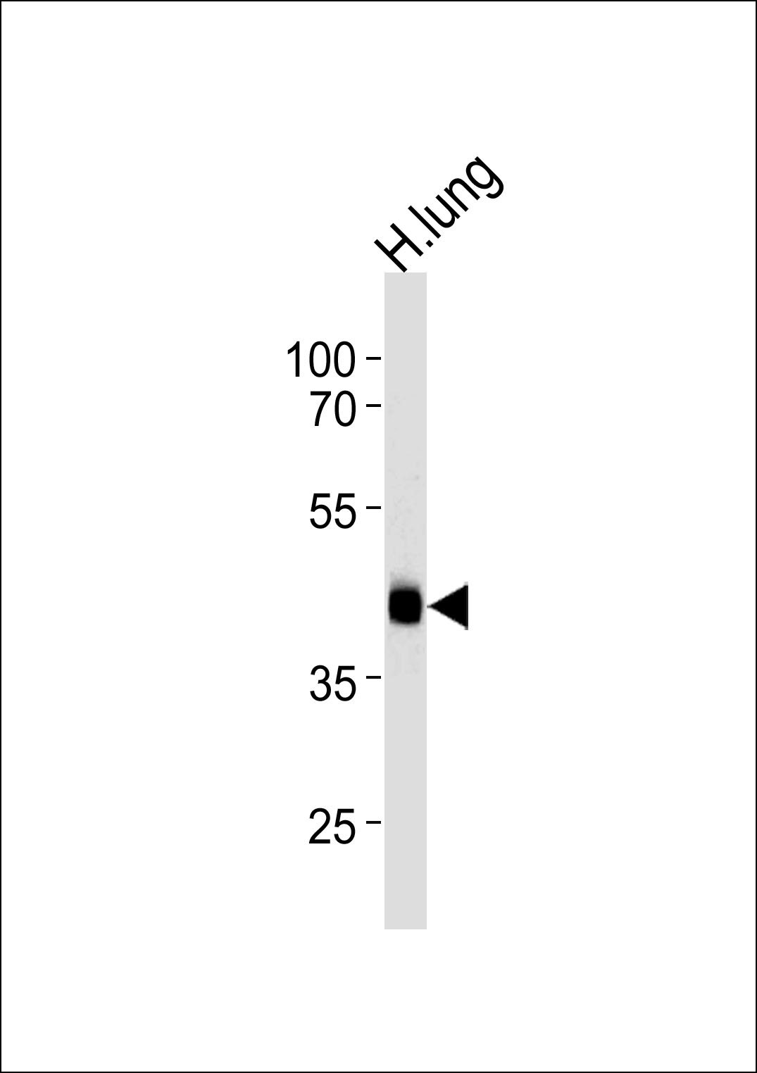
MTRF1 Antibody (Center)
Affinity Purified Rabbit Polyclonal Antibody (Pab)
- SPECIFICATION
- CITATIONS
- PROTOCOLS
- BACKGROUND

Application
| WB, E |
|---|---|
| Primary Accession | O75570 |
| Other Accession | Q8K126, Q3MHI7, NP_004285.2 |
| Reactivity | Human |
| Predicted | Bovine, Mouse |
| Host | Rabbit |
| Clonality | Polyclonal |
| Isotype | Rabbit IgG |
| Calculated MW | 52306 Da |
| Antigen Region | 232-259 aa |
| Gene ID | 9617 |
|---|---|
| Other Names | Peptide chain release factor 1, mitochondrial, MRF-1, MtRF-1, MTRF1 |
| Target/Specificity | This MTRF1 antibody is generated from rabbits immunized with a KLH conjugated synthetic peptide between 232-259 amino acids from the Central region of human MTRF1. |
| Dilution | WB~~1:1000 E~~Use at an assay dependent concentration. |
| Format | Purified polyclonal antibody supplied in PBS with 0.09% (W/V) sodium azide. This antibody is purified through a protein A column, followed by peptide affinity purification. |
| Storage | Maintain refrigerated at 2-8°C for up to 2 weeks. For long term storage store at -20°C in small aliquots to prevent freeze-thaw cycles. |
| Precautions | MTRF1 Antibody (Center) is for research use only and not for use in diagnostic or therapeutic procedures. |
| Name | RF1M |
|---|---|
| Function | Mitochondrial peptide chain release factor that directs the termination of translation in response to the peptide chain non- canonical stop codons AGG and AGA (PubMed:36302763, PubMed:36596788, PubMed:37141370). Non-canonical termination codons AGG and AGA are found at the end of MT-CO1/COX1 and MT-ND6/ND6 open reading frames, respectively (PubMed:37141370). Recognizes non-canonical stop codons via a network of interactions between the codon, MTRF1 and the ribosomal RNA (rRNA): in contrast to other translation release factors, which identify the codon in the A-site via direct interactions of amino acid side chains with the bases, MTRF1 repositions the first 2 bases of the stop codon to use an intricate network of interactions that includes residues of the release factor, the rRNA of the small ribosomal subunit, as well as neighboring bases of the mRNA (PubMed:37141370). |
| Cellular Location | Mitochondrion |

Thousands of laboratories across the world have published research that depended on the performance of antibodies from Abcepta to advance their research. Check out links to articles that cite our products in major peer-reviewed journals, organized by research category.
info@abcepta.com, and receive a free "I Love Antibodies" mug.

Provided below are standard protocols that you may find useful for product applications.
Background
The protein encoded by this gene was determined by in silico methods to be a mitochondrial protein with similarity to the peptide chain release factors (RFs) discovered in bacteria and yeast. The peptide chain release factors direct the termination of translation in response to the peptide chain termination codons. Initially thought to have a role in the termination of mitochondria protein synthesis, a recent publication found no mitochondrial translation release functionality. Multiple alternatively spliced transcript variants have been suggested by mRNA and EST data; however, their full-length natures are not clear. [provided by RefSeq].
References
Antonicka, H., et al. Am. J. Hum. Genet. 87(1):115-122(2010)
Nozaki, Y., et al. Genes Cells 13(5):429-438(2008)
Soleimanpour-Lichaei, H.R., et al. Mol. Cell 27(5):745-757(2007)
Hansen, L.L., et al. Cytogenet. Cell Genet. 88 (1-2), 91-92 (2000) :
Zhang, Y., et al. Biochim. Biophys. Acta 1443 (1-2), 245-250 (1998) :
If you have used an Abcepta product and would like to share how it has performed, please click on the "Submit Review" button and provide the requested information. Our staff will examine and post your review and contact you if needed.
If you have any additional inquiries please email technical services at tech@abcepta.com.
Ordering Information
Other Products
Shipping Information
