


 Foundational characteristics of cancer include proliferation, angiogenesis, migration, evasion of apoptosis, and cellular immortality. Find key markers for these cellular processes and antibodies to detect them.
 The SUMOplot™ Analysis Program predicts and scores sumoylation sites in your protein. SUMOylation is a post-translational modification involved in various cellular processes, such as nuclear-cytosolic transport, transcriptional regulation, apoptosis, protein stability, response to stress, and progression through the cell cycle.
 The Autophagy Receptor Motif Plotter predicts and scores autophagy receptor binding sites in your protein. Identifying proteins connected to this pathway is critical to understanding the role of autophagy in physiological as well as pathological processes such as development, differentiation, neurodegenerative diseases, stress, infection, and cancer.
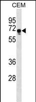
FEM1C Antibody (C-term)
Affinity Purified Rabbit Polyclonal Antibody (Pab)
- SPECIFICATION
- CITATIONS
- PROTOCOLS
- BACKGROUND

Application
| WB, E |
|---|---|
| Primary Accession | Q96JP0 |
| Other Accession | Q8CEF1, A7MB89, NP_064562.1 |
| Reactivity | Human |
| Predicted | Bovine, Mouse |
| Host | Rabbit |
| Clonality | Polyclonal |
| Isotype | Rabbit IgG |
| Calculated MW | 68673 Da |
| Antigen Region | 455-481 aa |
| Gene ID | 56929 |
|---|---|
| Other Names | Protein fem-1 homolog C, FEM1c, FEM1-gamma, FEM1C, KIAA1785 |
| Target/Specificity | This FEM1C antibody is generated from rabbits immunized with a KLH conjugated synthetic peptide between 455-481 amino acids from the C-terminal region of human FEM1C. |
| Dilution | WB~~1:1000 E~~Use at an assay dependent concentration. |
| Format | Purified polyclonal antibody supplied in PBS with 0.09% (W/V) sodium azide. This antibody is purified through a protein A column, followed by peptide affinity purification. |
| Storage | Maintain refrigerated at 2-8°C for up to 2 weeks. For long term storage store at -20°C in small aliquots to prevent freeze-thaw cycles. |
| Precautions | FEM1C Antibody (C-term) is for research use only and not for use in diagnostic or therapeutic procedures. |
| Name | FEM1C {ECO:0000303|PubMed:14527725, ECO:0000312|HGNC:HGNC:16933} |
|---|---|
| Function | Substrate-recognition component of a Cul2-RING (CRL2) E3 ubiquitin-protein ligase complex of the DesCEND (destruction via C-end degrons) pathway, which recognizes a C-degron located at the extreme C terminus of target proteins, leading to their ubiquitination and degradation (PubMed:29775578, PubMed:29779948, PubMed:33398168, PubMed:33398170, PubMed:38326650). The C-degron recognized by the DesCEND pathway is usually a motif of less than ten residues and can be present in full-length proteins, truncated proteins or proteolytically cleaved forms (PubMed:29775578, PubMed:29779948, PubMed:33398168, PubMed:33398170). The CRL2(FEM1C) complex specifically recognizes proteins with an arginine at the C-terminus: recognizes and binds proteins ending with -Lys/Arg-Xaa-Arg and -Lys/Arg-Xaa-Xaa-Arg C- degrons, such as SIL1 or OR51B2, leading to their ubiquitination and degradation (PubMed:33398168, PubMed:33398170, PubMed:38326650). The CRL2(FEM1C) complex mediates ubiquitination and degradation of truncated MSRB1/SEPX1 selenoproteins produced by failed UGA/Sec decoding (PubMed:26138980). Promotes ubiquitination and degradation of SLBP (PubMed:28118078). |
| Tissue Location | Widely expressed. Highly expressed in kidney, cardiac tissue, skeletal muscle and testis. Expressed at lower levels in other tissues, including cartilage. |

Thousands of laboratories across the world have published research that depended on the performance of antibodies from Abcepta to advance their research. Check out links to articles that cite our products in major peer-reviewed journals, organized by research category.
info@abcepta.com, and receive a free "I Love Antibodies" mug.

Provided below are standard protocols that you may find useful for product applications.
Background
Probable component of an E3 ubiquitin-protein ligase complex, in which it may act as a substrate recognition subunit (By similarity).
References
Goodarzi, M.O., et al. Hum. Reprod. 23(12):2842-2849(2008)
Ventura-Holman, T., et al. Gene 314, 133-139 (2003) :
Krakow, D., et al. Gene 279(2):213-219(2001)
If you have used an Abcepta product and would like to share how it has performed, please click on the "Submit Review" button and provide the requested information. Our staff will examine and post your review and contact you if needed.
If you have any additional inquiries please email technical services at tech@abcepta.com.
Ordering Information
Shipping Information
