


 Foundational characteristics of cancer include proliferation, angiogenesis, migration, evasion of apoptosis, and cellular immortality. Find key markers for these cellular processes and antibodies to detect them.
 The SUMOplot™ Analysis Program predicts and scores sumoylation sites in your protein. SUMOylation is a post-translational modification involved in various cellular processes, such as nuclear-cytosolic transport, transcriptional regulation, apoptosis, protein stability, response to stress, and progression through the cell cycle.
 The Autophagy Receptor Motif Plotter predicts and scores autophagy receptor binding sites in your protein. Identifying proteins connected to this pathway is critical to understanding the role of autophagy in physiological as well as pathological processes such as development, differentiation, neurodegenerative diseases, stress, infection, and cancer.
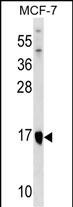
ITGB1BP1 Antibody (Center)
Affinity Purified Rabbit Polyclonal Antibody (Pab)
- SPECIFICATION
- CITATIONS
- PROTOCOLS
- BACKGROUND

Application
| WB, E |
|---|---|
| Primary Accession | O14713 |
| Other Accession | Q3ZBM4, NP_004754.1, NP_071729.1 |
| Reactivity | Human |
| Predicted | Bovine |
| Host | Rabbit |
| Clonality | Polyclonal |
| Isotype | Rabbit IgG |
| Calculated MW | 21782 Da |
| Antigen Region | 108-136 aa |
| Gene ID | 9270 |
|---|---|
| Other Names | Integrin beta-1-binding protein 1, Integrin cytoplasmic domain-associated protein 1, ICAP-1, ITGB1BP1, ICAP1 |
| Target/Specificity | This ITGB1BP1 antibody is generated from rabbits immunized with a KLH conjugated synthetic peptide between 108-136 amino acids from the Central region of human ITGB1BP1. |
| Dilution | WB~~1:1000 E~~Use at an assay dependent concentration. |
| Format | Purified polyclonal antibody supplied in PBS with 0.09% (W/V) sodium azide. This antibody is purified through a protein A column, followed by peptide affinity purification. |
| Storage | Maintain refrigerated at 2-8°C for up to 2 weeks. For long term storage store at -20°C in small aliquots to prevent freeze-thaw cycles. |
| Precautions | ITGB1BP1 Antibody (Center) is for research use only and not for use in diagnostic or therapeutic procedures. |
| Name | ITGB1BP1 |
|---|---|
| Synonyms | ICAP1 |
| Function | Key regulator of the integrin-mediated cell-matrix interaction signaling by binding to the ITGB1 cytoplasmic tail and preventing the activation of integrin alpha-5/beta-1 (heterodimer of ITGA5 and ITGB1) by talin or FERMT1. Plays a role in cell proliferation, differentiation, spreading, adhesion and migration in the context of mineralization and bone development and angiogenesis. Stimulates cellular proliferation in a fibronectin-dependent manner. Involved in the regulation of beta-1 integrin-containing focal adhesion (FA) site dynamics by controlling its assembly rate during cell adhesion; inhibits beta-1 integrin clustering within FA by directly competing with talin TLN1, and hence stimulates osteoblast spreading and migration in a fibronectin- and/or collagen-dependent manner. Acts as a guanine nucleotide dissociation inhibitor (GDI) by regulating Rho family GTPases during integrin-mediated cell matrix adhesion; reduces the level of active GTP-bound form of both CDC42 and RAC1 GTPases upon cell adhesion to fibronectin. Stimulates the release of active CDC42 from the membranes to maintain it in an inactive cytoplasmic pool. Participates in the translocation of the Rho-associated protein kinase ROCK1 to membrane ruffles at cell leading edges of the cell membrane, leading to an increase of myoblast cell migration on laminin. Plays a role in bone mineralization at a late stage of osteoblast differentiation; modulates the dynamic formation of focal adhesions into fibrillar adhesions, which are adhesive structures responsible for fibronectin deposition and fibrillogenesis. Plays a role in blood vessel development; acts as a negative regulator of angiogenesis by attenuating endothelial cell proliferation and migration, lumen formation and sprouting angiogenesis by promoting AKT phosphorylation and inhibiting ERK1/2 phosphorylation through activation of the Notch signaling pathway. Promotes transcriptional activity of the MYC promoter. |
| Cellular Location | Nucleus. Cytoplasm. Cytoplasm, cytoskeleton. Cell membrane. Cell projection, lamellipodium. Cell projection, ruffle. Note=Nucleocytoplasmic shuttling protein; shuttles between nucleus and cytoplasm in a integrin-dependent manner; probably sequestered in the cytosol by ITGB1. Its localization is dependent on the stage of cell spreading on fibronectin; cytoplasmic in case of round cells, corresponding to the initial step of cell spreading, or nuclear in case of well spread cells. Colocalizes with ROCK1 and NME2 at beta-1 integrin engagement sites. Together with ITGB1 and NME2 is recruited to beta-1 integrin- rich peripheral ruffles and lamellipodia during initial cell spreading on fibronectin and/or collagen |
| Tissue Location | Expressed in endothelial cells and fibroblasts (at protein level). Ubiquitously expressed. Expressed in intestine, colon, testis, ovary, thymus, spleen and prostate |

Thousands of laboratories across the world have published research that depended on the performance of antibodies from Abcepta to advance their research. Check out links to articles that cite our products in major peer-reviewed journals, organized by research category.
info@abcepta.com, and receive a free "I Love Antibodies" mug.

Provided below are standard protocols that you may find useful for product applications.
Background
The cytoplasmic domains of integrins are essential for cell adhesion. The protein encoded by this gene binds to the beta1 integrin cytoplasmic domain. The interaction between this protein and beta1 integrin is highly specific. Two isoforms of this protein are derived from alternatively spliced transcripts. The shorter form of this protein does not interact with the beta1 integrin cytoplasmic domain. The longer form is a phosphoprotein and the extent of its phosphorylation is regulated by the cell-matrix interaction, suggesting an important role of this protein during integrin-dependent cell adhesion.
References
Brutsch, R., et al. Circ. Res. 107(5):592-601(2010)
Mavaddat, N., et al. Cancer Epidemiol. Biomarkers Prev. 18(1):255-259(2009)
Zhang, J., et al. Neurosurgery 63(3):571-578(2008)
Furusu, A., et al. J. Cell. Physiol. 210(3):703-710(2007)
Stroeken, P.J., et al. J. Cell. Physiol. 208(3):620-628(2006)
If you have used an Abcepta product and would like to share how it has performed, please click on the "Submit Review" button and provide the requested information. Our staff will examine and post your review and contact you if needed.
If you have any additional inquiries please email technical services at tech@abcepta.com.
Ordering Information
Other Products
Shipping Information
